6RU8

Crystal structure of Casein Kinase I delta (CK1d) in complex with triple phosphorylated p63 PAD3P peptide


Experimental Data Snapshot

  • Method: X-RAY DIFFRACTION
  • Resolution: 1.92 Å
  • R-Value Free: 0.211 
  • R-Value Work: 0.172 
  • R-Value Observed: 0.174 

wwPDB Validation   3D Report Full Report


Ligand Structure Quality Assessment 


This is version 1.3 of the entry. See complete history


Literature

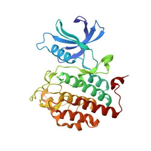
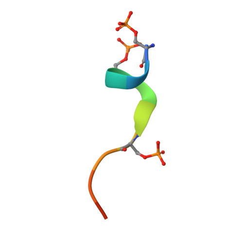
p63 uses a switch-like mechanism to set the threshold for induction of apoptosis.

Gebel, J.Tuppi, M.Chaikuad, A.Hotte, K.Schroder, M.Schulz, L.Lohr, F.Gutfreund, N.Finke, F.Henrich, E.Mezhyrova, J.Lehnert, R.Pampaloni, F.Hummer, G.Stelzer, E.H.K.Knapp, S.Dotsch, V.

(2020) Nat Chem Biol 16: 1078-1086

  • DOI: https://doi.org/10.1038/s41589-020-0600-3
  • Primary Citation of Related Structures:  
    6RU6, 6RU7, 6RU8

  • PubMed Abstract: 

    The p53 homolog TAp63α is the transcriptional key regulator of genome integrity in oocytes. After DNA damage, TAp63α is activated by multistep phosphorylation involving multiple phosphorylation events by the kinase CK1, which triggers the transition from a dimeric and inactive conformation to an open and active tetramer that initiates apoptosis. By measuring activation kinetics in ovaries and single-site phosphorylation kinetics in vitro with peptides and full-length protein, we show that TAp63α phosphorylation follows a biphasic behavior. Although the first two CK1 phosphorylation events are fast, the third one, which constitutes the decisive step to form the active conformation, is slow. Structure determination of CK1 in complex with differently phosphorylated peptides reveals the structural mechanism for the difference in the kinetic behavior based on an unusual CK1/TAp63α substrate interaction in which the product of one phosphorylation step acts as an inhibitor for the following one.


  • Organizational Affiliation

    Institute of Biophysical Chemistry and Center for Biomolecular Magnetic Resonance and Cluster of Excellence Macromolecular Complexes (CEF), Goethe University, Frankfurt am Main, Germany.


Macromolecules
Find similar proteins by:  (by identity cutoff)  |  3D Structure
Entity ID: 1
MoleculeChains Sequence LengthOrganismDetailsImage
Casein kinase I isoform delta
A, B, C, D
296Homo sapiensMutation(s): 0 
Gene Names: CSNK1DHCKID
EC: 2.7.11.1 (PDB Primary Data), 2.7.11.26 (PDB Primary Data)
UniProt & NIH Common Fund Data Resources
Find proteins for P48730 (Homo sapiens)
Explore P48730 
Go to UniProtKB:  P48730
PHAROS:  P48730
GTEx:  ENSG00000141551 
Entity Groups  
Sequence Clusters30% Identity50% Identity70% Identity90% Identity95% Identity100% Identity
UniProt GroupP48730
Sequence Annotations
Expand
  • Reference Sequence

Find similar proteins by:  Sequence   |   3D Structure  

Entity ID: 2
MoleculeChains Sequence LengthOrganismDetailsImage
Tumor protein 63
E, F, G, H
12Homo sapiensMutation(s): 0 
UniProt & NIH Common Fund Data Resources
Find proteins for Q9H3D4 (Homo sapiens)
Explore Q9H3D4 
Go to UniProtKB:  Q9H3D4
PHAROS:  Q9H3D4
GTEx:  ENSG00000073282 
Entity Groups  
Sequence Clusters30% Identity50% Identity70% Identity90% Identity95% Identity100% Identity
UniProt GroupQ9H3D4
Sequence Annotations
Expand
  • Reference Sequence
Small Molecules
Ligands 4 Unique
IDChains Name / Formula / InChI Key2D Diagram3D Interactions
ADP
Query on ADP

Download Ideal Coordinates CCD File 
FA [auth D],
I [auth A],
Q [auth B],
Y [auth C]
ADENOSINE-5'-DIPHOSPHATE
C10 H15 N5 O10 P2
XTWYTFMLZFPYCI-KQYNXXCUSA-N
SO4
Query on SO4

Download Ideal Coordinates CCD File 
P [auth A],
W [auth B],
X [auth B]
SULFATE ION
O4 S
QAOWNCQODCNURD-UHFFFAOYSA-L
EDO
Query on EDO

Download Ideal Coordinates CCD File 
AA [auth C]
BA [auth C]
CA [auth C]
DA [auth C]
EA [auth C]
AA [auth C],
BA [auth C],
CA [auth C],
DA [auth C],
EA [auth C],
GA [auth D],
HA [auth D],
IA [auth D],
J [auth A],
K [auth A],
L [auth A],
M [auth A],
N [auth A],
R [auth B],
S [auth B],
T [auth B],
U [auth B],
Z [auth C]
1,2-ETHANEDIOL
C2 H6 O2
LYCAIKOWRPUZTN-UHFFFAOYSA-N
NA
Query on NA

Download Ideal Coordinates CCD File 
O [auth A],
V [auth B]
SODIUM ION
Na
FKNQFGJONOIPTF-UHFFFAOYSA-N
Modified Residues  1 Unique
IDChains TypeFormula2D DiagramParent
SEP
Query on SEP
E, F, G, H
L-PEPTIDE LINKINGC3 H8 N O6 PSER
Experimental Data & Validation

Experimental Data

  • Method: X-RAY DIFFRACTION
  • Resolution: 1.92 Å
  • R-Value Free: 0.211 
  • R-Value Work: 0.172 
  • R-Value Observed: 0.174 
  • Space Group: P 1
Unit Cell:
Length ( Å )Angle ( ˚ )
a = 48.727α = 108.6
b = 84.131β = 105.83
c = 89.287γ = 91.57
Software Package:
Software NamePurpose
Aimlessdata scaling
REFMACrefinement
PDB_EXTRACTdata extraction
XDSdata reduction
PHASERphasing

Structure Validation

View Full Validation Report


Ligand Structure Quality Assessment 


Entry History 

Revision History  (Full details and data files)

  • Version 1.0: 2020-05-13
    Type: Initial release
  • Version 1.1: 2020-08-12
    Changes: Database references, Derived calculations
  • Version 1.2: 2020-09-30
    Changes: Database references
  • Version 1.3: 2024-01-24
    Changes: Data collection, Database references, Refinement description