6RRM

Crystal structure of LdtMt2 from Mycobacterium tuberculosis bound to Ebselen


Experimental Data Snapshot

  • Method: X-RAY DIFFRACTION
  • Resolution: 1.64 Å
  • R-Value Free: 0.223 
  • R-Value Work: 0.200 
  • R-Value Observed: 0.201 

wwPDB Validation   3D Report Full Report


Ligand Structure Quality Assessment 


This is version 1.4 of the entry. See complete history


Literature

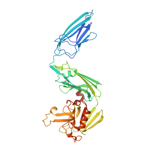
Targeting the Mycobacterium tuberculosis transpeptidase LdtMt2with cysteine-reactive inhibitors including ebselen.

de Munnik, M.Lohans, C.T.Lang, P.A.Langley, G.W.Malla, T.R.Tumber, A.Schofield, C.J.Brem, J.

(2019) Chem Commun (Camb) 55: 10214-10217

  • DOI: https://doi.org/10.1039/c9cc04145a
  • Primary Citation of Related Structures:  
    6RLG, 6RRM

  • PubMed Abstract: 

    The l,d-transpeptidases (Ldts) are promising antibiotic targets for treating tuberculosis. We report screening of cysteine-reactive inhibitors against LdtMt2 from Mycobacterium tuberculosis. Structural studies on LdtMt2 with potent inhibitor ebselen reveal opening of the benzisoselenazolone ring by a nucleophilic cysteine, forming a complex involving extensive hydrophobic interactions with a substrate-binding loop.


  • Organizational Affiliation

    Chemistry Research Laboratory, Department of Chemistry, University of Oxford, Oxford, OX1 3TA, UK. christopher.schofield@chem.ox.ac.uk jurgen.brem@chem.ox.ac.uk.


Macromolecules
Find similar proteins by:  (by identity cutoff)  |  3D Structure
Entity ID: 1
MoleculeChains Sequence LengthOrganismDetailsImage
L,D-transpeptidase 2
A, B
355Mycobacterium tuberculosis CDC1551Mutation(s): 0 
Gene Names: ldtBMT2594V735_02606
EC: 2.3.2
UniProt
Find proteins for I6Y9J2 (Mycobacterium tuberculosis (strain ATCC 25618 / H37Rv))
Explore I6Y9J2 
Go to UniProtKB:  I6Y9J2
Entity Groups  
Sequence Clusters30% Identity50% Identity70% Identity90% Identity95% Identity100% Identity
UniProt GroupI6Y9J2
Sequence Annotations
Expand
  • Reference Sequence
Small Molecules
Ligands 4 Unique
IDChains Name / Formula / InChI Key2D Diagram3D Interactions
9JT (Subject of Investigation/LOI)
Query on 9JT

Download Ideal Coordinates CCD File 
F [auth A],
I [auth B]
N-phenyl-2-selanylbenzamide
C13 H11 N O Se
PVPUYGNPKBMXGO-UHFFFAOYSA-N
GOL
Query on GOL

Download Ideal Coordinates CCD File 
D [auth A]GLYCEROL
C3 H8 O3
PEDCQBHIVMGVHV-UHFFFAOYSA-N
EDO
Query on EDO

Download Ideal Coordinates CCD File 
G [auth B],
J [auth B]
1,2-ETHANEDIOL
C2 H6 O2
LYCAIKOWRPUZTN-UHFFFAOYSA-N
NH4
Query on NH4

Download Ideal Coordinates CCD File 
C [auth A],
E [auth A],
H [auth B]
AMMONIUM ION
H4 N
QGZKDVFQNNGYKY-UHFFFAOYSA-O
Experimental Data & Validation

Experimental Data

  • Method: X-RAY DIFFRACTION
  • Resolution: 1.64 Å
  • R-Value Free: 0.223 
  • R-Value Work: 0.200 
  • R-Value Observed: 0.201 
  • Space Group: P 1 21 1
Unit Cell:
Length ( Å )Angle ( ˚ )
a = 61.22α = 90
b = 93.57β = 93.01
c = 75.49γ = 90
Software Package:
Software NamePurpose
XDSdata reduction
Aimlessdata scaling
PHASERphasing
PHENIXrefinement
PDB_EXTRACTdata extraction

Structure Validation

View Full Validation Report


Ligand Structure Quality Assessment 


Entry History & Funding Information

Deposition Data


Funding OrganizationLocationGrant Number
Medical Research Council (United Kingdom)United Kingdom--

Revision History  (Full details and data files)

  • Version 1.0: 2019-08-14
    Type: Initial release
  • Version 1.1: 2019-08-21
    Changes: Data collection, Derived calculations
  • Version 1.2: 2019-09-04
    Changes: Data collection, Database references
  • Version 1.3: 2023-04-12
    Changes: Database references, Derived calculations, Structure summary
  • Version 1.4: 2024-02-07
    Changes: Data collection, Refinement description