6RP5

Crystal structure of monocarboxylated hemoglobin from the sub-Antarctic fish Eleginops maclovinus


Experimental Data Snapshot

  • Method: X-RAY DIFFRACTION
  • Resolution: 1.49 Å
  • R-Value Free: 0.196 
  • R-Value Work: 0.168 
  • R-Value Observed: 0.170 

wwPDB Validation   3D Report Full Report


Ligand Structure Quality Assessment 


This is version 1.1 of the entry. See complete history


Literature

The unique structural features of carbonmonoxy hemoglobin from the sub-Antarctic fish Eleginops maclovinus.

Balasco, N.Vitagliano, L.Merlino, A.Verde, C.Mazzarella, L.Vergara, A.

(2019) Sci Rep 9: 18987-18987

  • DOI: https://doi.org/10.1038/s41598-019-55331-3
  • Primary Citation of Related Structures:  
    6RP5

  • PubMed Abstract: 

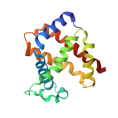
    Tetrameric hemoglobins (Hbs) are prototypical systems for the investigations of fundamental properties of proteins. Although the structure of these proteins has been known for nearly sixty years, there are many aspects related to their function/structure that are still obscure. Here, we report the crystal structure of a carbonmonoxy form of the Hb isolated from the sub-Antarctic notothenioid fish Eleginops maclovinus characterised by either rare or unique features. In particular, the distal site of the α chain results to be very unusual since the distal His is displaced from its canonical position. This displacement is coupled with a shortening of the highly conserved E helix and the formation of novel interactions at tertiary structure level. Interestingly, the quaternary structure is closer to the T-deoxy state of Hbs than to the R-state despite the full coordination of all chains. Notably, these peculiar structural features provide a rationale for some spectroscopic properties exhibited by the protein in solution. Finally, this unexpected structural plasticity of the heme distal side has been associated with specific sequence signatures of various Hbs.


  • Organizational Affiliation

    Institute of Biostructures and Bioimaging, CNR, Via Mezzocannone 16, Naples, Italy.


Macromolecules
Find similar proteins by:  (by identity cutoff)  |  3D Structure
Entity ID: 1
MoleculeChains Sequence LengthOrganismDetailsImage
Hemoglobin subunit alpha 1143Eleginops maclovinusMutation(s): 1 
UniProt
Find proteins for K7N5M5 (Eleginops maclovinus)
Explore K7N5M5 
Go to UniProtKB:  K7N5M5
Entity Groups  
Sequence Clusters30% Identity50% Identity70% Identity90% Identity95% Identity100% Identity
UniProt GroupK7N5M5
Sequence Annotations
Expand
  • Reference Sequence
Find similar proteins by:  (by identity cutoff)  |  3D Structure
Entity ID: 2
MoleculeChains Sequence LengthOrganismDetailsImage
Hemoglobin subunit beta-1144Eleginops maclovinusMutation(s): 0 
UniProt
Find proteins for K7N5M6 (Eleginops maclovinus)
Explore K7N5M6 
Go to UniProtKB:  K7N5M6
Entity Groups  
Sequence Clusters30% Identity50% Identity70% Identity90% Identity95% Identity100% Identity
UniProt GroupK7N5M6
Sequence Annotations
Expand
  • Reference Sequence
Small Molecules
Ligands 4 Unique
IDChains Name / Formula / InChI Key2D Diagram3D Interactions
HEM
Query on HEM

Download Ideal Coordinates CCD File 
D [auth A],
I [auth B]
PROTOPORPHYRIN IX CONTAINING FE
C34 H32 Fe N4 O4
KABFMIBPWCXCRK-RGGAHWMASA-L
DTN
Query on DTN

Download Ideal Coordinates CCD File 
G [auth A]DITHIONITE
O4 S2
GRWZHXKQBITJKP-UHFFFAOYSA-L
SO4
Query on SO4

Download Ideal Coordinates CCD File 
E [auth A],
F [auth A],
J [auth B],
K [auth B]
SULFATE ION
O4 S
QAOWNCQODCNURD-UHFFFAOYSA-L
CMO
Query on CMO

Download Ideal Coordinates CCD File 
C [auth A],
H [auth B]
CARBON MONOXIDE
C O
UGFAIRIUMAVXCW-UHFFFAOYSA-N
Experimental Data & Validation

Experimental Data

  • Method: X-RAY DIFFRACTION
  • Resolution: 1.49 Å
  • R-Value Free: 0.196 
  • R-Value Work: 0.168 
  • R-Value Observed: 0.170 
  • Space Group: P 61 2 2
Unit Cell:
Length ( Å )Angle ( ˚ )
a = 91.702α = 90
b = 91.702β = 90
c = 168.716γ = 120
Software Package:
Software NamePurpose
REFMACrefinement
HKL-2000data reduction
HKL-2000data scaling
PHASERphasing

Structure Validation

View Full Validation Report


Ligand Structure Quality Assessment 


Entry History 

Deposition Data

Revision History  (Full details and data files)

  • Version 1.0: 2019-12-25
    Type: Initial release
  • Version 1.1: 2024-01-24
    Changes: Data collection, Database references, Refinement description