6RNL

L-[Ru(TAP)2(dppz)]2+ bound to the G-quadruplex forming sequence d(TAGGGTT)


Experimental Data Snapshot

  • Method: X-RAY DIFFRACTION
  • Resolution: 1.88 Å
  • R-Value Free: 0.214 
  • R-Value Work: 0.187 
  • R-Value Observed: 0.189 

wwPDB Validation   3D Report Full Report


Ligand Structure Quality Assessment 


This is version 1.1 of the entry. See complete history


Literature

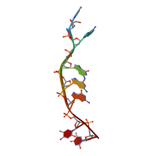
Three thymine/adenine binding modes of the ruthenium complex Lambda-[Ru(TAP)2(dppz)]2+to the G-quadruplex forming sequence d(TAGGGTT) shown by X-ray crystallography.

McQuaid, K.Hall, J.P.Baumgaertner, L.Cardin, D.J.Cardin, C.J.

(2019) Chem Commun (Camb) 55: 9116-9119

  • DOI: https://doi.org/10.1039/c9cc04316k
  • Primary Citation of Related Structures:  
    6RNL

  • PubMed Abstract: 

    Λ-[Ru(TAP)2(dppz)]2+ was crystallised with the G-quadruplex-forming heptamer d(TAGGGTT). Surprisingly, even though there are four unique binding sites, the complex is not in contact with any G-quartet surface. Two complexes stabilise cavities formed from terminal T·A and T·T mismatched pairs. A third shows kinking by a TAP ligand between T·T linkages, while the fourth shows sandwiching of a dppz ligand between a T·A/T·A quadruplex and a T·T mismatch, stabilised by an additional T·A base pair stacking interaction on a TAP surface. Overall, the structure shows an unexpected affinity for thymine, and suggests models for G-quadruplex loop binding.


  • Organizational Affiliation

    Department of Chemistry, University of Reading, Whiteknights, Reading, RG6 6AD, UK. c.j.cardin@reading.ac.uk.


Macromolecules

Find similar nucleic acids by:  Sequence   |   3D Structure  

Entity ID: 1
MoleculeChains LengthOrganismImage
DNA (5'-D(*TP*AP*GP*GP*GP*TP*T)-3')
A, B, C, D
7synthetic construct
Sequence Annotations
Expand
  • Reference Sequence
Experimental Data & Validation

Experimental Data

  • Method: X-RAY DIFFRACTION
  • Resolution: 1.88 Å
  • R-Value Free: 0.214 
  • R-Value Work: 0.187 
  • R-Value Observed: 0.189 
  • Space Group: P 65
Unit Cell:
Length ( Å )Angle ( ˚ )
a = 38.514α = 90
b = 38.514β = 90
c = 128.804γ = 120
Software Package:
Software NamePurpose
PHENIXrefinement
DIALSdata reduction
DIALSdata scaling
SHELXDEphasing
xia2data scaling

Structure Validation

View Full Validation Report


Ligand Structure Quality Assessment 


Entry History & Funding Information

Deposition Data


Funding OrganizationLocationGrant Number
Biotechnology and Biological Sciences Research CouncilUnited KingdomBB/K019279/1
Biotechnology and Biological Sciences Research CouncilUnited KingdomBB/M004635/1

Revision History  (Full details and data files)

  • Version 1.0: 2019-08-28
    Type: Initial release
  • Version 1.1: 2019-09-11
    Changes: Data collection, Database references