6RKS

E. coli DNA Gyrase - DNA binding and cleavage domain in State 1 without TOPRIM insertion


Experimental Data Snapshot

  • Method: ELECTRON MICROSCOPY
  • Resolution: 4.00 Å
  • Aggregation State: PARTICLE 
  • Reconstruction Method: SINGLE PARTICLE 

wwPDB Validation   3D Report Full Report


This is version 1.3 of the entry. See complete history


Literature

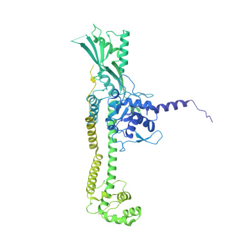
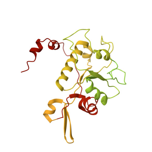
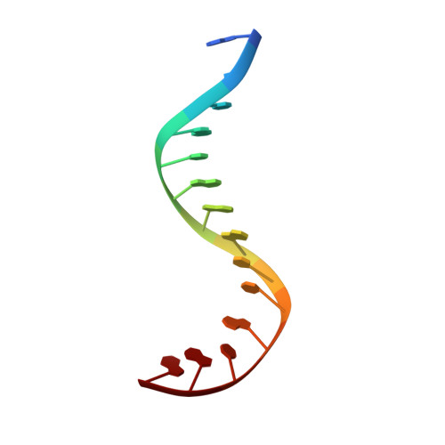
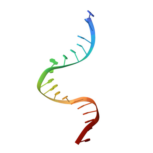
Cryo-EM structure of the complete E. coli DNA gyrase nucleoprotein complex.

Vanden Broeck, A.Lotz, C.Ortiz, J.Lamour, V.

(2019) Nat Commun 10: 4935-4935

  • DOI: https://doi.org/10.1038/s41467-019-12914-y
  • Primary Citation of Related Structures:  
    6RKS, 6RKU, 6RKV, 6RKW

  • PubMed Abstract: 

    DNA gyrase is an essential enzyme involved in the homeostatic control of DNA supercoiling and the target of successful antibacterial compounds. Despite extensive studies, a detailed architecture of the full-length DNA gyrase from the model organism E. coli is still missing. Herein, we report the complete structure of the E. coli DNA gyrase nucleoprotein complex trapped by the antibiotic gepotidacin, using phase-plate single-particle cryo-electron microscopy. Our data unveil the structural and spatial organization of the functional domains, their connections and the position of the conserved GyrA-box motif. The deconvolution of two states of the DNA-binding/cleavage domain provides a better understanding of the allosteric movements of the enzyme complex. The local atomic resolution in the DNA-bound area reaching up to 3.0 Å enables the identification of the antibiotic density. Altogether, this study paves the way for the cryo-EM determination of gyrase complexes with antibiotics and opens perspectives for targeting conformational intermediates.


  • Organizational Affiliation

    Department of Integrated Structural Biology, Institut de Génétique et de Biologie Moléculaire et Cellulaire (IGBMC), 1 Rue Laurent Fries, 67404, Illkirch Cedex, France.


Macromolecules

Find similar proteins by:  (by identity cutoff)  |  3D Structure
Entity ID: 1
MoleculeChains Sequence LengthOrganismDetailsImage
DNA gyrase subunit A
A, C
875Escherichia coli K-12Mutation(s): 0 
Gene Names: gyrAhisWnalAparDb2231JW2225
EC: 5.6.2.2
UniProt
Find proteins for P0AES4 (Escherichia coli (strain K12))
Explore P0AES4 
Go to UniProtKB:  P0AES4
Entity Groups  
Sequence Clusters30% Identity50% Identity70% Identity90% Identity95% Identity100% Identity
UniProt GroupP0AES4
Sequence Annotations
Expand
  • Reference Sequence
Find similar proteins by:  (by identity cutoff)  |  3D Structure
Entity ID: 2
MoleculeChains Sequence LengthOrganismDetailsImage
DNA gyrase subunit B
B, D
804Escherichia coli K-12Mutation(s): 0 
Gene Names: gyrBacrBcouhimBhisUnalCparApcbAb3699JW5625
EC: 5.6.2.2
UniProt
Find proteins for P0AES6 (Escherichia coli (strain K12))
Explore P0AES6 
Go to UniProtKB:  P0AES6
Entity Groups  
Sequence Clusters30% Identity50% Identity70% Identity90% Identity95% Identity100% Identity
UniProt GroupP0AES6
Sequence Annotations
Expand
  • Reference Sequence

Find similar nucleic acids by:  Sequence   |   3D Structure  

Entity ID: 3
MoleculeChains LengthOrganismImage
DNA Strand 1E [auth F],
F [auth E]
14Escherichia coli
Sequence Annotations
Expand
  • Reference Sequence

Find similar nucleic acids by:  Sequence   |   3D Structure  

Entity ID: 4
MoleculeChains LengthOrganismImage
DNA Strand 2
G, H
18Escherichia coli
Sequence Annotations
Expand
  • Reference Sequence
Small Molecules
Ligands 1 Unique
IDChains Name / Formula / InChI Key2D Diagram3D Interactions
JHN
Query on JHN

Download Ideal Coordinates CCD File 
I [auth G](3~{R})-3-[[4-(3,4-dihydro-2~{H}-pyrano[2,3-c]pyridin-6-ylmethylamino)piperidin-1-yl]methyl]-1,4,7-triazatricyclo[6.3.1.0^{4,12}]dodeca-6,8(12),9-triene-5,11-dione
C24 H28 N6 O3
PZFAZQUREQIODZ-LJQANCHMSA-N
Experimental Data & Validation

Experimental Data

  • Method: ELECTRON MICROSCOPY
  • Resolution: 4.00 Å
  • Aggregation State: PARTICLE 
  • Reconstruction Method: SINGLE PARTICLE 
EM Software:
TaskSoftware PackageVersion
RECONSTRUCTIONRELION2.0.3
MODEL REFINEMENTPHENIX1.12

Structure Validation

View Full Validation Report


Entry History & Funding Information

Deposition Data


Funding OrganizationLocationGrant Number
French National Research AgencyFranceANR-10-LABX-0030-INRT
French National Research AgencyFranceANR-10-IDEX-0002-02
French Infrastructure for Integrated Structural BiologyFranceFRISBI ANR-10-INBS-05

Revision History  (Full details and data files)

  • Version 1.0: 2019-11-06
    Type: Initial release
  • Version 1.1: 2019-11-13
    Changes: Data collection, Database references
  • Version 1.2: 2020-02-05
    Changes: Database references, Source and taxonomy, Structure summary
  • Version 1.3: 2020-11-18
    Changes: Data collection