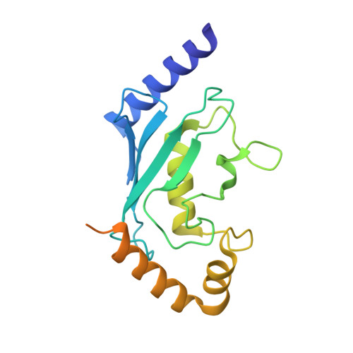
6R75

Crystal structure of human Ube2T E54R mutant


Experimental Data Snapshot

  • Method: X-RAY DIFFRACTION
  • Resolution: 2.00 Å
  • R-Value Free: 0.264 
  • R-Value Work: 0.225 
  • R-Value Observed: 0.227 

wwPDB Validation   3D Report Full Report


This is version 1.3 of the entry. See complete history


Literature

Allosteric mechanism for site-specific ubiquitination of FANCD2.

Chaugule, V.K.Arkinson, C.Rennie, M.L.Kamarainen, O.Toth, R.Walden, H.

(2020) Nat Chem Biol 16: 291-301

  • DOI: https://doi.org/10.1038/s41589-019-0426-z
  • Primary Citation of Related Structures:  
    6R75

  • PubMed Abstract: 

    DNA-damage repair is implemented by proteins that are coordinated by specialized molecular signals. One such signal in the Fanconi anemia (FA) pathway for the repair of DNA interstrand crosslinks is the site-specific monoubiquitination of FANCD2 and FANCI. The signal is mediated by a multiprotein FA core complex (FA-CC) however, the mechanics for precise ubiquitination remain elusive. We show that FANCL, the RING-bearing module in FA-CC, allosterically activates its cognate ubiqutin-conjugating enzyme E2 UBE2T to drive site-specific FANCD2 ubiquitination. Unlike typical RING E3 ligases, FANCL catalyzes ubiquitination by rewiring the intraresidue network of UBE2T to influence the active site. Consequently, a basic triad unique to UBE2T engages a structured acidic patch near the target lysine on FANCD2. This three-dimensional complementarity, between the E2 active site and substrate surface, induced by FANCL is central to site-specific monoubiquitination in the FA pathway. Furthermore, the allosteric network of UBE2T can be engineered to enhance FANCL-catalyzed FANCD2-FANCI di-monoubiquitination without compromising site specificity.


  • Organizational Affiliation

    Institute of Molecular, Cell and Systems Biology, College of Medical, Veterinary and Life Sciences, University of Glasgow, Glasgow, UK. Chaugule.Viduth@gmail.com.


Macromolecules
Find similar proteins by:  (by identity cutoff)  |  3D Structure
Entity ID: 1
MoleculeChains Sequence LengthOrganismDetailsImage
Ubiquitin-conjugating enzyme E2 T201Homo sapiensMutation(s): 1 
Gene Names: UBE2THSPC150PIG50
EC: 2.3.2.23
UniProt & NIH Common Fund Data Resources
Find proteins for Q9NPD8 (Homo sapiens)
Explore Q9NPD8 
Go to UniProtKB:  Q9NPD8
PHAROS:  Q9NPD8
GTEx:  ENSG00000077152 
Entity Groups  
Sequence Clusters30% Identity50% Identity70% Identity90% Identity95% Identity100% Identity
UniProt GroupQ9NPD8
Sequence Annotations
Expand
  • Reference Sequence
Experimental Data & Validation

Experimental Data

  • Method: X-RAY DIFFRACTION
  • Resolution: 2.00 Å
  • R-Value Free: 0.264 
  • R-Value Work: 0.225 
  • R-Value Observed: 0.227 
  • Space Group: P 21 21 21
Unit Cell:
Length ( Å )Angle ( ˚ )
a = 46.13α = 90
b = 46.19β = 90
c = 74.2γ = 90
Software Package:
Software NamePurpose
MOSFLMdata reduction
Aimlessdata scaling
PHASERphasing
REFMACrefinement
PDB_EXTRACTdata extraction

Structure Validation

View Full Validation Report


Entry History & Funding Information

Deposition Data


Funding OrganizationLocationGrant Number
Medical Research Council (United Kingdom)United KingdomMC_UU_12016/12
European Research CouncilUnited KingdomERC-2015-CoG-681582

Revision History  (Full details and data files)

  • Version 1.0: 2019-10-16
    Type: Initial release
  • Version 1.1: 2020-01-22
    Changes: Database references
  • Version 1.2: 2020-03-04
    Changes: Database references
  • Version 1.3: 2024-01-24
    Changes: Data collection, Database references, Refinement description