


Crystal structure of the periplasmic nickel-binding protein NikA from Escherichia coli in complex with Ru(bpza)CO H2O Cl
Cavazza, C., Menage, S.To be published.
Experimental Data Snapshot
Starting Model: experimental
View more details
Entity ID: 1 | |||||
|---|---|---|---|---|---|
| Molecule | Chains | Sequence Length | Organism | Details | Image |
| Nickel-binding periplasmic protein | 502 | Escherichia coli K-12 | Mutation(s): 0 Gene Names: nikA, b3476, JW3441 | 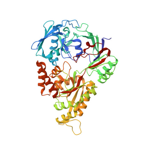 | |
UniProt | |||||
Entity Groups | |||||
| Sequence Clusters | 30% Identity50% Identity70% Identity90% Identity95% Identity100% Identity | ||||
| UniProt Group | P33590 | ||||
Sequence AnnotationsExpand | |||||
Reference Sequence | |||||
| Ligands 8 Unique | |||||
|---|---|---|---|---|---|
| ID | Chains | Name / Formula / InChI Key | 2D Diagram | 3D Interactions | |
| EDT Download:Ideal Coordinates CCD File | D [auth A] | {[-(BIS-CARBOXYMETHYL-AMINO)-ETHYL]-CARBOXYMETHYL-AMINO}-ACETIC ACID C10 H16 N2 O8 KCXVZYZYPLLWCC-UHFFFAOYSA-N |  | ||
| 6RP Download:Ideal Coordinates CCD File | X [auth B] | bis(pyrzol-1-yl)acetate scorpionate C8 H8 N4 O2 NJDSSVBTTVUKHM-UHFFFAOYSA-N |  | ||
| RU Download:Ideal Coordinates CCD File | W [auth B] | RUTHENIUM ION Ru BPEVHDGLPIIAGH-UHFFFAOYSA-N |  | ||
| GOL Download:Ideal Coordinates CCD File | AA [auth B] BA [auth B] CA [auth B] DA [auth B] EA [auth B] | GLYCEROL C3 H8 O3 PEDCQBHIVMGVHV-UHFFFAOYSA-N |  | ||
| ACT Download:Ideal Coordinates CCD File | GA [auth B] HA [auth B] M [auth A] N [auth A] O [auth A] | ACETATE ION C2 H3 O2 QTBSBXVTEAMEQO-UHFFFAOYSA-M |  | ||
| FE Download:Ideal Coordinates CCD File | C [auth A] | FE (III) ION Fe VTLYFUHAOXGGBS-UHFFFAOYSA-N |  | ||
| CL Download:Ideal Coordinates CCD File | E [auth A], Y [auth B] | CHLORIDE ION Cl VEXZGXHMUGYJMC-UHFFFAOYSA-M |  | ||
| CMO Download:Ideal Coordinates CCD File | Z [auth B] | CARBON MONOXIDE C O UGFAIRIUMAVXCW-UHFFFAOYSA-N |  | ||
| Length ( Å ) | Angle ( ˚ ) |
|---|---|
| a = 86.629 | α = 90 |
| b = 94.101 | β = 90 |
| c = 124.418 | γ = 90 |
| Software Name | Purpose |
|---|---|
| REFMAC | refinement |
| XDS | data reduction |
| XSCALE | data scaling |
| REFMAC | phasing |
| Funding Organization | Location | Grant Number |
|---|---|---|
| French National Research Agency | France | Crystalball |


