6R2N

Crystal structure of KlGlk1 glucokinase from Kluyveromyces lactis


Experimental Data Snapshot

  • Method: X-RAY DIFFRACTION
  • Resolution: 2.60 Å
  • R-Value Free: 0.243 
  • R-Value Work: 0.204 
  • R-Value Observed: 0.205 

wwPDB Validation   3D Report Full Report


This is version 1.2 of the entry. See complete history


Literature

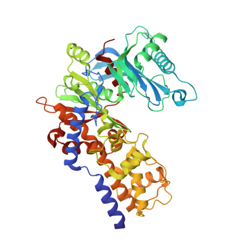
Crystal Structure of Kluyveromyces lactis Glucokinase ( Kl Glk1).

Zak, K.M.Kalinska, M.Wator, E.Kuska, K.Krutyholowa, R.Dubin, G.Popowicz, G.M.Grudnik, P.

(2019) Int J Mol Sci 20

  • DOI: https://doi.org/10.3390/ijms20194821
  • Primary Citation of Related Structures:  
    6R2N

  • PubMed Abstract: 

    Glucose phosphorylating enzymes are crucial in the regulation of basic cellular processes, including metabolism and gene expression. Glucokinases and hexokinases provide a pool of phosphorylated glucose in an adenosine diphosphate (ADP)- and ATP-dependent manner to shape the cell metabolism. The glucose processing enzymes from Kluyveromyces lactis are poorly characterized despite the emerging contribution of this yeast strain to industrial and laboratory scale biotechnology. The first reports on K. lactis glucokinase ( Kl Glk1) positioned the enzyme as an essential component required for glucose signaling. Nevertheless, no biochemical and structural information was available until now. Here, we present the first crystal structure of Kl Glk1 together with biochemical characterization, including substrate specificity and enzyme kinetics. Additionally, comparative analysis of the presented structure and the prior structures of lactis hexokinase ( Kl Hxk1) demonstrates the potential transitions between open and closed enzyme conformations upon ligand binding.


  • Organizational Affiliation

    Institute of Structural Biology, Helmholtz Zentrum München, Ingolstädter Landstrasse 1, 85764 Neuherberg, Germany. krzysztof.zak@helmholtz-muenchen.de.


Macromolecules
Find similar proteins by:  (by identity cutoff)  |  3D Structure
Entity ID: 1
MoleculeChains Sequence LengthOrganismDetailsImage
Glucokinase-1
A, B, C
475Kluyveromyces lactisMutation(s): 0 
Gene Names: GLK1KLLA0C01155g
EC: 2.7.1.2
UniProt
Find proteins for Q6CUZ3 (Kluyveromyces lactis (strain ATCC 8585 / CBS 2359 / DSM 70799 / NBRC 1267 / NRRL Y-1140 / WM37))
Explore Q6CUZ3 
Go to UniProtKB:  Q6CUZ3
Entity Groups  
Sequence Clusters30% Identity50% Identity70% Identity90% Identity95% Identity100% Identity
UniProt GroupQ6CUZ3
Sequence Annotations
Expand
  • Reference Sequence
Small Molecules
Ligands 2 Unique
IDChains Name / Formula / InChI Key2D Diagram3D Interactions
BR
Query on BR

Download Ideal Coordinates CCD File 
O [auth C]BROMIDE ION
Br
CPELXLSAUQHCOX-UHFFFAOYSA-M
EDO
Query on EDO

Download Ideal Coordinates CCD File 
D [auth A]
E [auth A]
F [auth A]
G [auth A]
H [auth A]
D [auth A],
E [auth A],
F [auth A],
G [auth A],
H [auth A],
I [auth A],
J [auth A],
K [auth A],
L [auth B],
M [auth B],
N [auth B]
1,2-ETHANEDIOL
C2 H6 O2
LYCAIKOWRPUZTN-UHFFFAOYSA-N
Experimental Data & Validation

Experimental Data

  • Method: X-RAY DIFFRACTION
  • Resolution: 2.60 Å
  • R-Value Free: 0.243 
  • R-Value Work: 0.204 
  • R-Value Observed: 0.205 
  • Space Group: C 2 2 21
Unit Cell:
Length ( Å )Angle ( ˚ )
a = 102.011α = 90
b = 122.771β = 90
c = 360.346γ = 90
Software Package:
Software NamePurpose
PHENIXrefinement
PDB_EXTRACTdata extraction
XDSdata reduction
XDSdata scaling
PHASERphasing

Structure Validation

View Full Validation Report


Entry History & Funding Information

Deposition Data


Funding OrganizationLocationGrant Number
Polish National Science CentrePolandUMO-2015/19/D/NZ1/02009

Revision History  (Full details and data files)

  • Version 1.0: 2019-10-16
    Type: Initial release
  • Version 1.1: 2020-09-30
    Changes: Database references
  • Version 1.2: 2024-01-24
    Changes: Advisory, Data collection, Database references, Refinement description