6R02

Psychrobacter arcticus ATP phosphoribosyltransferase bound to histidine and PRPP


Experimental Data Snapshot

  • Method: X-RAY DIFFRACTION
  • Resolution: 2.65 Å
  • R-Value Free: 0.270 
  • R-Value Work: 0.236 
  • R-Value Observed: 0.238 

Starting Model: experimental
View more details

wwPDB Validation   3D Report Full Report


Ligand Structure Quality Assessment 


This is version 1.2 of the entry. See complete history


Literature

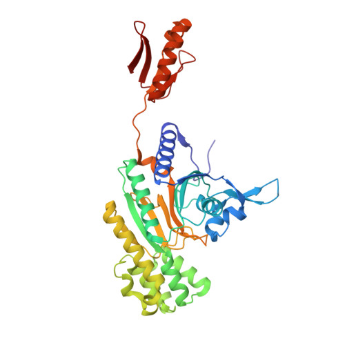
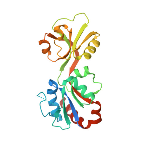
Mapping the Structural Path for Allosteric Inhibition of a Short-Form ATP Phosphoribosyltransferase by Histidine.

Thomson, C.M.Alphey, M.S.Fisher, G.da Silva, R.G.

(2019) Biochemistry 58: 3078-3086

  • DOI: https://doi.org/10.1021/acs.biochem.9b00282
  • Primary Citation of Related Structures:  
    6R02

  • PubMed Abstract: 

    ATP phosphoribosyltransferase (ATPPRT) catalyzes the first step of histidine biosynthesis, being allosterically inhibited by the final product of the pathway. Allosteric inhibition of long-form ATPPRTs by histidine has been extensively studied, but inhibition of short-form ATPPRTs is poorly understood. Short-form ATPPRTs are hetero-octamers formed by four catalytic subunits (HisG S ) and four regulatory subunits (HisZ). HisG S alone is catalytically active and insensitive to histidine. HisZ enhances catalysis by HisG S in the absence of histidine but mediates allosteric inhibition in its presence. Here, steady-state and pre-steady-state kinetics establish that histidine is a noncompetitive inhibitor of short-form ATPPRT and that inhibition does not occur by dissociating HisG S from the hetero-octamer. The crystal structure of ATPPRT in complex with histidine and the substrate 5-phospho-α-d-ribosyl-1-pyrophosphate was determined, showing histidine bound solely to HisZ, with four histidine molecules per hetero-octamer. Histidine binding involves the repositioning of two HisZ loops. The histidine-binding loop moves closer to histidine to establish polar contacts. This leads to a hydrogen bond between its Tyr263 and His104 in the Asp101-Leu117 loop. The Asp101-Leu117 loop leads to the HisZ-HisG S interface, and in the absence of histidine, its motion prompts HisG S conformational changes responsible for catalytic activation. Following histidine binding, interaction with the histidine-binding loop may prevent the Asp101-Leu117 loop from efficiently sampling conformations conducive to catalytic activation. Tyr263Phe- Pa HisZ-containing Pa ATPPRT, however, is less susceptible though not insensitive to histidine inhibition, suggesting the Tyr263-His104 interaction may be relevant to yet not solely responsible for transmission of the allosteric signal.


  • Organizational Affiliation

    School of Biology, Biomedical Sciences Research Complex , University of St Andrews , St Andrews , Fife KY16 9ST , U.K.


Macromolecules
Find similar proteins by:  (by identity cutoff)  |  3D Structure
Entity ID: 1
MoleculeChains Sequence LengthOrganismDetailsImage
ATP phosphoribosyltransferase regulatory subunit
A, B, C, D
388Psychrobacter arcticusMutation(s): 0 
Gene Names: hisZPsyc_0676
UniProt
Find proteins for Q4FTX3 (Psychrobacter arcticus (strain DSM 17307 / VKM B-2377 / 273-4))
Explore Q4FTX3 
Go to UniProtKB:  Q4FTX3
Entity Groups  
Sequence Clusters30% Identity50% Identity70% Identity90% Identity95% Identity100% Identity
UniProt GroupQ4FTX3
Sequence Annotations
Expand
  • Reference Sequence
Find similar proteins by:  (by identity cutoff)  |  3D Structure
Entity ID: 2
MoleculeChains Sequence LengthOrganismDetailsImage
ATP phosphoribosyltransferase
E, F, G, H
232Psychrobacter arcticusMutation(s): 0 
Gene Names: hisGPsyc_1903
EC: 2.4.2.17
UniProt
Find proteins for Q4FQF7 (Psychrobacter arcticus (strain DSM 17307 / VKM B-2377 / 273-4))
Explore Q4FQF7 
Go to UniProtKB:  Q4FQF7
Entity Groups  
Sequence Clusters30% Identity50% Identity70% Identity90% Identity95% Identity100% Identity
UniProt GroupQ4FQF7
Sequence Annotations
Expand
  • Reference Sequence
Experimental Data & Validation

Experimental Data

  • Method: X-RAY DIFFRACTION
  • Resolution: 2.65 Å
  • R-Value Free: 0.270 
  • R-Value Work: 0.236 
  • R-Value Observed: 0.238 
  • Space Group: P 1 21 1
Unit Cell:
Length ( Å )Angle ( ˚ )
a = 93.253α = 90
b = 147.31β = 103.11
c = 99.327γ = 90
Software Package:
Software NamePurpose
REFMACrefinement
XDSdata reduction
Aimlessdata scaling
MOLREPphasing
PDB_EXTRACTdata extraction

Structure Validation

View Full Validation Report


Ligand Structure Quality Assessment 


Entry History 

Deposition Data

Revision History  (Full details and data files)

  • Version 1.0: 2019-08-07
    Type: Initial release
  • Version 1.1: 2020-07-29
    Type: Remediation
    Reason: Carbohydrate remediation
    Changes: Data collection, Derived calculations, Structure summary
  • Version 1.2: 2024-01-24
    Changes: Data collection, Database references, Refinement description, Structure summary