6QXA | pdb_00006qxa

Structure of membrane bound pyrophosphatase from Thermotoga maritima in complex with imidodiphosphate and N-[(2-amino-6-benzothiazolyl)methyl]-1H-indole-2-carboxamide (ATC)


Experimental Data Snapshot

  • Method: X-RAY DIFFRACTION
  • Resolution: 3.41 Å
  • R-Value Free: 
    0.284 (Depositor), 0.286 (DCC) 
  • R-Value Work: 
    0.228 (Depositor), 0.231 (DCC) 
  • R-Value Observed: 
    0.231 (Depositor) 

Starting Model: experimental
View more details

wwPDB Validation 3D Report Full Report

Validation slider image for 6QXA

Ligand Structure Quality Assessment 


This is version 1.4 of the entry. See complete history

Re-refinement Note

This entry reflects an alternative modeling of the original data in: 6HT3

Literature

Asymmetry in catalysis byThermotoga maritimamembrane-bound pyrophosphatase demonstrated by a nonphosphorus allosteric inhibitor.

Vidilaseris, K.Kiriazis, A.Turku, A.Khattab, A.Johansson, N.G.Leino, T.O.Kiuru, P.S.Boije Af Gennas, G.Meri, S.Yli-Kauhaluoma, J.Xhaard, H.Goldman, A.

(2019) Sci Adv 5: eaav7574-eaav7574

  • DOI: https://doi.org/10.1126/sciadv.aav7574
  • Primary Citation Related Structures: 
    6QXA

  • PubMed Abstract: 

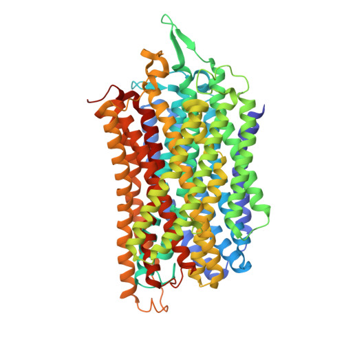
    Membrane-bound pyrophosphatases are homodimeric integral membrane proteins that hydrolyze pyrophosphate into orthophosphates, coupled to the active transport of protons or sodium ions across membranes. They are important in the life cycle of bacteria, archaea, plants, and parasitic protists, but no homologous proteins exist in vertebrates, making them a promising drug target. Here, we report the first nonphosphorus allosteric inhibitor of the thermophilic bacterium Thermotoga maritima membrane-bound pyrophosphatase and its bound structure together with the substrate analog imidodiphosphate. The unit cell contains two protein homodimers, each binding a single inhibitor dimer near the exit channel, creating a hydrophobic clamp that inhibits the movement of β-strand 1-2 during pumping, and thus prevents the hydrophobic gate from opening. This asymmetry of inhibitor binding with respect to each homodimer provides the first clear structural demonstration of asymmetry in the catalytic cycle of membrane-bound pyrophosphatases.


  • Organizational Affiliation
    • Research Program in Molecular and Integrative Biosciences, University of Helsinki, Helsinki, Finland.

Macromolecule Content 

  • Total Structure Weight: 315.37 kDa 
  • Atom Count: 21,042 
  • Modeled Residue Count: 2,884 
  • Deposited Residue Count: 2,940 
  • Unique protein chains: 1

Macromolecules

Find similar proteins by:|  3D Structure
Entity ID: 1
MoleculeChains  Sequence LengthOrganismDetailsImage
K(+)-stimulated pyrophosphate-energized sodium pump
A, B, C, D
735Thermotoga maritima MSB8Mutation(s): 0 
Gene Names: hppATM_0174
EC: 7.2.3.1
Membrane Entity: Yes 
UniProt
Find proteins for Q9S5X0 (Thermotoga maritima (strain ATCC 43589 / DSM 3109 / JCM 10099 / NBRC 100826 / MSB8))
Explore Q9S5X0 
Go to UniProtKB:  Q9S5X0
Entity Groups
Sequence Clusters30% Identity50% Identity70% Identity90% Identity95% Identity100% Identity
UniProt GroupQ9S5X0
Sequence Annotations
Expand
Reference Sequence

Small Molecules

Ligands 6 Unique
IDChains Name / Formula / InChI Key2D Diagram3D Interactions
GQB

Query on GQB


Download:Ideal Coordinates CCD File
DA [auth C],
M [auth A],
N [auth A]
~{N}-[(2-azanyl-3~{H}-1,3-benzothiazol-6-yl)methyl]-1~{H}-indole-2-carboxamide
C17 H16 N4 O S
GAUGHKFDIIXTPX-UHFFFAOYSA-N
2PN

Query on 2PN


Download:Ideal Coordinates CCD File
BA [auth C],
J [auth A],
JA [auth D],
T [auth B]
IMIDODIPHOSPHORIC ACID
H5 N O6 P2
GNGSOPFGGKKDQP-UHFFFAOYSA-N
D1D

Query on D1D


Download:Ideal Coordinates CCD File
CA [auth C](4S,5S)-1,2-DITHIANE-4,5-DIOL
C4 H8 O2 S2
YPGMOWHXEQDBBV-QWWZWVQMSA-N
K

Query on K


Download:Ideal Coordinates CCD File
L [auth A],
LA [auth D],
V [auth B]
POTASSIUM ION
K
NPYPAHLBTDXSSS-UHFFFAOYSA-N
MG

Query on MG


Download:Ideal Coordinates CCD File
AA [auth C]
E [auth A]
EA [auth D]
F [auth A]
FA [auth D]
AA [auth C],
E [auth A],
EA [auth D],
F [auth A],
FA [auth D],
G [auth A],
GA [auth D],
H [auth A],
HA [auth D],
I [auth A],
IA [auth D],
O [auth B],
P [auth B],
Q [auth B],
R [auth B],
S [auth B],
W [auth C],
X [auth C],
Y [auth C],
Z [auth C]
MAGNESIUM ION
Mg
JLVVSXFLKOJNIY-UHFFFAOYSA-N
NA

Query on NA


Download:Ideal Coordinates CCD File
K [auth A],
KA [auth D],
U [auth B]
SODIUM ION
Na
FKNQFGJONOIPTF-UHFFFAOYSA-N

Experimental Data & Validation

Experimental Data

  • Method: X-RAY DIFFRACTION
  • Resolution: 3.41 Å
  • R-Value Free:  0.284 (Depositor), 0.286 (DCC) 
  • R-Value Work:  0.228 (Depositor), 0.231 (DCC) 
  • R-Value Observed: 0.231 (Depositor) 
Space Group: P 21 21 21
Unit Cell:
Length ( Å )Angle ( ˚ )
a = 98.101α = 90
b = 141.594β = 90
c = 251.98γ = 90
Software Package:
Software NamePurpose
PHENIXrefinement
XDSdata reduction
Aimlessdata scaling
PHASERphasing

Structure Validation

View Full Validation Report


Ligand Structure Quality Assessment 


Entry History 

& Funding Information

Deposition Data


Funding OrganizationLocationGrant Number
Academy of FinlandFinland308105
Academy of FinlandFinland310297
Academy of FinlandFinland265481
Academy of FinlandFinland279755
Biotechnology and Biological Sciences Research CouncilUnited KingdomBB/M021610
Jane and Aatos Erkko FoundationFinland--

Revision History  (Full details and data files)

  • Version 1.0: 2019-04-10
    Type: Initial release
  • Version 1.1: 2019-05-15
    Changes: Advisory, Data collection, Database references
  • Version 1.2: 2019-06-05
    Changes: Data collection, Database references
  • Version 1.3: 2023-03-08
    Changes: Advisory, Database references, Derived calculations
  • Version 1.4: 2024-02-07
    Changes: Data collection, Refinement description