6QU9 | pdb_00006qu9

Fab fragment of an antibody that inhibits polymerisation of alpha-1-antitrypsin


Experimental Data Snapshot

  • Method: X-RAY DIFFRACTION
  • Resolution: 1.90 Å
  • R-Value Free: 
    0.233 (Depositor), 0.236 (DCC) 
  • R-Value Work: 
    0.202 (Depositor), 0.204 (DCC) 
  • R-Value Observed: 
    0.203 (Depositor) 

Starting Model: other
View more details

wwPDB Validation 3D Report Full Report

Validation slider image for 6QU9

This is version 1.3 of the entry. See complete history

Literature

The structural basis for Z alpha 1 -antitrypsin polymerization in the liver.

Faull, S.V.Elliston, E.L.K.Gooptu, B.Jagger, A.M.Aldobiyan, I.Redzej, A.Badaoui, M.Heyer-Chauhan, N.Rashid, S.T.Reynolds, G.M.Adams, D.H.Miranda, E.Orlova, E.V.Irving, J.A.Lomas, D.A.

(2020) Sci Adv 6

  • DOI: https://doi.org/10.1126/sciadv.abc1370
  • Primary Citation Related Structures: 
    6QU9

  • PubMed Abstract: 

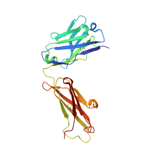
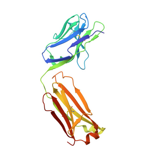
    The serpinopathies are among a diverse set of conformational diseases that involve the aberrant self-association of proteins into ordered aggregates. α 1 -Antitrypsin deficiency is the archetypal serpinopathy and results from the formation and deposition of mutant forms of α 1 -antitrypsin as "polymer" chains in liver tissue. No detailed structural analysis has been performed of this material. Moreover, there is little information on the relevance of well-studied artificially induced polymers to these disease-associated molecules. We have isolated polymers from the liver tissue of Z α 1 -antitrypsin homozygotes (E342K) who have undergone transplantation, labeled them using a Fab fragment, and performed single-particle analysis of negative-stain electron micrographs. The data show structural equivalence between heat-induced and ex vivo polymers and that the intersubunit linkage is best explained by a carboxyl-terminal domain swap between molecules of α 1 -antitrypsin.


  • Organizational Affiliation
    • UCL Respiratory, University College London, 5 University Street, London WC1E 6JF, UK.

Macromolecule Content 

  • Total Structure Weight: 94.12 kDa 
  • Atom Count: 6,885 
  • Modeled Residue Count: 848 
  • Deposited Residue Count: 854 
  • Unique protein chains: 2

Macromolecules

Find similar proteins by:|  3D Structure
Entity ID: 1
MoleculeChains  Sequence LengthOrganismDetailsImage
FAB 4B12 heavy chainA [auth H],
C [auth A]
215Mus musculusMutation(s): 0 
Find similar proteins by:|  3D Structure
Entity ID: 2
MoleculeChains  Sequence LengthOrganismDetailsImage
FAB 4B12 light chainB [auth L],
D [auth B]
212Mus musculusMutation(s): 0 

Experimental Data & Validation

Experimental Data

  • Method: X-RAY DIFFRACTION
  • Resolution: 1.90 Å
  • R-Value Free:  0.233 (Depositor), 0.236 (DCC) 
  • R-Value Work:  0.202 (Depositor), 0.204 (DCC) 
  • R-Value Observed: 0.203 (Depositor) 
Space Group: P 21 21 21
Unit Cell:
Length ( Å )Angle ( ˚ )
a = 79.268α = 90
b = 105.102β = 90
c = 105.128γ = 90
Software Package:
Software NamePurpose
PHENIXrefinement
XDSdata reduction
Aimlessdata scaling
PHASERphasing
PDB_EXTRACTdata extraction

Structure Validation

View Full Validation Report


Entry History 

& Funding Information

Deposition Data


Funding OrganizationLocationGrant Number
Medical Research Council (MRC, United Kingdom)United KingdomMR/N024842/1
Other governmentUnited KingdomUCLH/NIHR Biomedical Research Centre
Other privateUnited StatesAlpha-1 Foundation project grant to J. Irving

Revision History  (Full details and data files)

  • Version 1.0: 2020-03-18
    Type: Initial release
  • Version 1.1: 2020-11-04
    Changes: Database references
  • Version 1.2: 2024-05-01
    Changes: Data collection, Database references, Refinement description
  • Version 1.3: 2024-11-20
    Changes: Structure summary