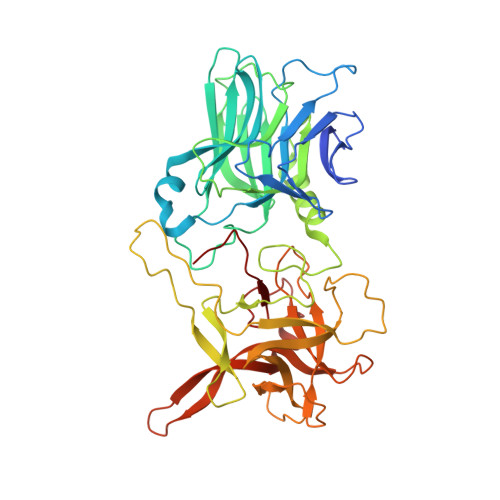
6QNS

Crystal structure of the binding domain of Botulinum Neurotoxin type B mutant I1248W/V1249W in complex with human synaptotagmin 1 and GD1a receptors


Experimental Data Snapshot

  • Method: X-RAY DIFFRACTION
  • Resolution: 2.40 Å
  • R-Value Free: 0.229 
  • R-Value Work: 0.199 
  • R-Value Observed: 0.201 

Starting Model: experimental
View more details

wwPDB Validation   3D Report Full Report


This is version 2.1 of the entry. See complete history


Literature

Characterization of a membrane binding loop leads to engineering botulinum neurotoxin B with improved therapeutic efficacy.

Yin, L.Masuyer, G.Zhang, S.Zhang, J.Miyashita, S.I.Burgin, D.Lovelock, L.Coker, S.F.Fu, T.M.Stenmark, P.Dong, M.

(2020) PLoS Biol 18: e3000618-e3000618

  • DOI: https://doi.org/10.1371/journal.pbio.3000618
  • Primary Citation of Related Structures:  
    6QNS

  • PubMed Abstract: 

    Botulinum neurotoxins (BoNTs) are a family of bacterial toxins with seven major serotypes (BoNT/A-G). The ability of these toxins to target and bind to motor nerve terminals is a key factor determining their potency and efficacy. Among these toxins, BoNT/B is one of the two types approved for medical and cosmetic uses. Besides binding to well-established receptors, an extended loop in the C-terminal receptor-binding domain (HC) of BoNT/B (HC/B) has been proposed to also contribute to toxin binding to neurons by interacting with lipid membranes (termed lipid-binding loop [LBL]). Analogous loops exist in the HCs of BoNT/C, D, G, and a chimeric toxin DC. However, it has been challenging to detect and characterize binding of LBLs to lipid membranes. Here, using the nanodisc system and biolayer interferometry assays, we find that HC/DC, C, and G, but not HC/B and HC/D, are capable of binding to receptor-free lipids directly, with HC/DC having the highest level of binding. Mutagenesis studies demonstrate the critical role of consecutive aromatic residues at the tip of the LBL for binding of HC/DC to lipid membranes. Taking advantage of this insight, we then create a "gain-of-function" mutant HC/B by replacing two nonaromatic residues at the tip of its LBL with tryptophan. Cocrystallization studies confirm that these two tryptophan residues do not alter the structure of HC/B or the interactions with its receptors. Such a mutated HC/B gains the ability to bind receptor-free lipid membranes and shows enhanced binding to cultured neurons. Finally, full-length BoNT/B containing two tryptophan mutations in its LBL, together with two additional mutations (E1191M/S1199Y) that increase binding to human receptors, is produced and evaluated in mice in vivo using Digit Abduction Score assays. This mutant toxin shows enhanced efficacy in paralyzing local muscles at the injection site and lower systemic diffusion, thus extending both safety range and duration of paralysis compared with the control BoNT/B. These findings establish a mechanistic understanding of LBL-lipid interactions and create a modified BoNT/B with improved therapeutic efficacy.


  • Organizational Affiliation

    Department of Urology, Boston Children's Hospital, Department of Microbiology and Department of Surgery, Harvard Medical School, Boston, Massachusetts, United States of America.


Macromolecules
Find similar proteins by:  (by identity cutoff)  |  3D Structure
Entity ID: 1
MoleculeChains Sequence LengthOrganismDetailsImage
Botulinum neurotoxin type B442Clostridium botulinumMutation(s): 2 
Gene Names: botB
EC: 3.4.24.69
UniProt
Find proteins for P10844 (Clostridium botulinum)
Explore P10844 
Go to UniProtKB:  P10844
Entity Groups  
Sequence Clusters30% Identity50% Identity70% Identity90% Identity95% Identity100% Identity
UniProt GroupP10844
Sequence Annotations
Expand
  • Reference Sequence

Find similar proteins by:  Sequence   |   3D Structure  

Entity ID: 2
MoleculeChains Sequence LengthOrganismDetailsImage
Synaptotagmin-1B [auth S]21Homo sapiensMutation(s): 0 
UniProt & NIH Common Fund Data Resources
Find proteins for P21579 (Homo sapiens)
Explore P21579 
Go to UniProtKB:  P21579
PHAROS:  P21579
GTEx:  ENSG00000067715 
Entity Groups  
Sequence Clusters30% Identity50% Identity70% Identity90% Identity95% Identity100% Identity
UniProt GroupP21579
Sequence Annotations
Expand
  • Reference Sequence
Oligosaccharides

Help

Entity ID: 3
MoleculeChains Length2D Diagram Glycosylation3D Interactions
N-acetyl-alpha-neuraminic acid-(2-3)-beta-D-galactopyranose-(1-3)-2-acetamido-2-deoxy-beta-D-galactopyranose-(1-4)-[N-acetyl-alpha-neuraminic acid-(2-3)]beta-D-galactopyranose-(1-4)-beta-D-glucopyranoseC [auth B]6N/A
Glycosylation Resources
GlyTouCan:  G46677TE
GlyCosmos:  G46677TE
GlyGen:  G46677TE
Experimental Data & Validation

Experimental Data

  • Method: X-RAY DIFFRACTION
  • Resolution: 2.40 Å
  • R-Value Free: 0.229 
  • R-Value Work: 0.199 
  • R-Value Observed: 0.201 
  • Space Group: P 21 21 21
Unit Cell:
Length ( Å )Angle ( ˚ )
a = 50.344α = 90
b = 78.491β = 90
c = 149.103γ = 90
Software Package:
Software NamePurpose
REFMACrefinement
DIALSdata reduction
Aimlessdata scaling
PHASERphasing

Structure Validation

View Full Validation Report


Entry History & Funding Information

Deposition Data


Funding OrganizationLocationGrant Number
Swedish Research CouncilSweden--

Revision History  (Full details and data files)

  • Version 1.0: 2020-02-26
    Type: Initial release
  • Version 1.1: 2020-03-25
    Changes: Database references
  • Version 2.0: 2020-07-29
    Type: Remediation
    Reason: Carbohydrate remediation
    Changes: Advisory, Atomic model, Data collection, Derived calculations, Structure summary
  • Version 2.1: 2024-01-24
    Changes: Data collection, Database references, Derived calculations, Refinement description, Structure summary