6QJO | pdb_00006qjo

DNA containing both right- and left-handed parallel-stranded G-quadruplexes


Experimental Data Snapshot

  • Method: X-RAY DIFFRACTION
  • Resolution: 1.80 Å
  • R-Value Free: 
    0.202 (Depositor), 0.196 (DCC) 
  • R-Value Work: 
    0.164 (Depositor), 0.170 (DCC) 
  • R-Value Observed: 
    0.172 (Depositor) 

Starting Model: experimental
View more details

wwPDB Validation 3D Report Full Report

Validation slider image for 6QJO

This is version 1.2 of the entry. See complete history

Literature

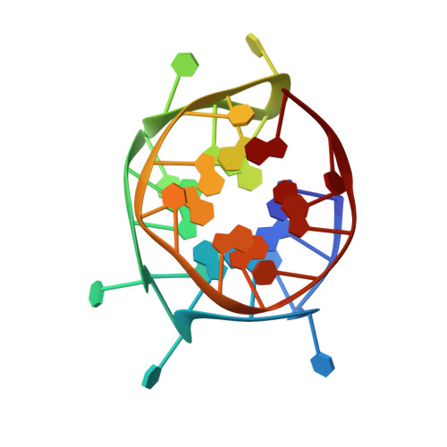
NMR solution and X-ray crystal structures of a DNA molecule containing both right- and left-handed parallel-stranded G-quadruplexes.

Winnerdy, F.R.Bakalar, B.Maity, A.Vandana, J.J.Mechulam, Y.Schmitt, E.Phan, A.T.

(2019) Nucleic Acids Res 47: 8272-8281

  • DOI: https://doi.org/10.1093/nar/gkz349
  • Primary Citation Related Structures: 
    6JCE, 6QJO

  • PubMed Abstract: 

    Analogous to the B- and Z-DNA structures in double-helix DNA, there exist both right- and left-handed quadruple-helix (G-quadruplex) DNA. Numerous conformations of right-handed and a few left-handed G-quadruplexes were previously observed, yet they were always identified separately. Here, we present the NMR solution and X-ray crystal structures of a right- and left-handed hybrid G-quadruplex. The structure reveals a stacking interaction between two G-quadruplex blocks with different helical orientations and displays features of both right- and left-handed G-quadruplexes. An analysis of loop mutations suggests that single-nucleotide loops are preferred or even required for the left-handed G-quadruplex formation. The discovery of a right- and left-handed hybrid G-quadruplex further expands the polymorphism of G-quadruplexes and is potentially useful in designing a left-to-right junction in G-quadruplex engineering.


  • Organizational Affiliation
    • School of Physical and Mathematical Sciences, Nanyang Technological University, Singapore 637371, Singapore.

Macromolecule Content 

  • Total Structure Weight: 36.18 kDa 
  • Atom Count: 2,545 
  • Modeled Residue Count: 112 
  • Deposited Residue Count: 112 
  • Unique nucleic acid chains: 1

Macromolecules

Find similar nucleic acids by:  (by identity cutoff) 
Entity ID: 1
MoleculeChains LengthOrganismImage
DNA (28-MER)
A, B, C, D
28synthetic construct
Sequence Annotations
Expand
Reference Sequence

Small Molecules

Ligands 1 Unique
IDChains Name / Formula / InChI Key2D Diagram3D Interactions
K

Query on K


Download:Ideal Coordinates CCD File
E [auth A]
F [auth A]
G [auth A]
H [auth A]
I [auth B]
E [auth A],
F [auth A],
G [auth A],
H [auth A],
I [auth B],
J [auth B],
K [auth B],
L [auth C],
M [auth C],
N [auth C],
O [auth C],
P [auth D],
Q [auth D],
R [auth D],
S [auth D]
POTASSIUM ION
K
NPYPAHLBTDXSSS-UHFFFAOYSA-N

Experimental Data & Validation

Experimental Data

  • Method: X-RAY DIFFRACTION
  • Resolution: 1.80 Å
  • R-Value Free:  0.202 (Depositor), 0.196 (DCC) 
  • R-Value Work:  0.164 (Depositor), 0.170 (DCC) 
  • R-Value Observed: 0.172 (Depositor) 
Space Group: P 1
Unit Cell:
Length ( Å )Angle ( ˚ )
a = 31.12α = 90.04
b = 39.27β = 93.03
c = 53.55γ = 89.95
Software Package:
Software NamePurpose
PHENIXrefinement
XDSdata reduction
XSCALEdata scaling
PHASERphasing

Structure Validation

View Full Validation Report


Entry History 

Deposition Data

Revision History  (Full details and data files)

  • Version 1.0: 2019-07-03
    Type: Initial release
  • Version 1.1: 2019-09-18
    Changes: Data collection, Database references
  • Version 1.2: 2024-01-24
    Changes: Data collection, Database references, Derived calculations, Refinement description