Tying up the Loose Ends: A Mathematically Knotted Protein.
Hsu, S.D., Lee, Y.C., Mikula, K.M., Backlund, S.M., Tascon, I., Goldman, A., Iwai, H.(2021) Front Chem 9: 663241-663241
- PubMed: 34109153 Search on PubMedSearch on PubMed Central
- DOI: https://doi.org/10.3389/fchem.2021.663241
- Primary Citation Related Structures:
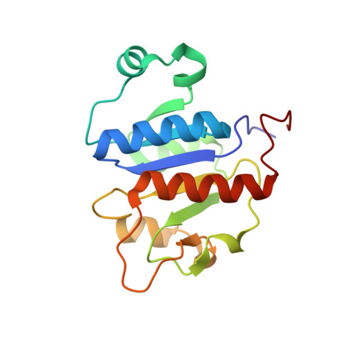
6QH8, 6QKV - PubMed Abstract:
Knots have attracted scientists in mathematics, physics, biology, and engineering. Long flexible thin strings easily knot and tangle as experienced in our daily life. Similarly, long polymer chains inevitably tend to get trapped into knots. Little is known about their formation or function in proteins despite >1,000 knotted proteins identified in nature. However, these protein knots are not mathematical knots with their backbone polypeptide chains because of their open termini, and the presence of a "knot" depends on the algorithm used to create path closure. Furthermore, it is generally not possible to control the topology of the unfolded states of proteins, therefore making it challenging to characterize functional and physicochemical properties of knotting in any polymer. Covalently linking the amino and carboxyl termini of the deeply trefoil-knotted YibK from Pseudomonas aeruginosa allowed us to create the truly backbone knotted protein by enzymatic peptide ligation. Moreover, we produced and investigated backbone cyclized YibK without any knotted structure. Thus, we could directly probe the effect of the backbone knot and the decrease in conformational entropy on protein folding. The backbone cyclization did not perturb the native structure and its cofactor binding affinity, but it substantially increased the thermal stability and reduced the aggregation propensity. The enhanced stability of a backbone knotted YibK could be mainly originated from an increased ruggedness of its free energy landscape and the destabilization of the denatured state by backbone cyclization with little contribution from a knot structure. Despite the heterogeneity in the side-chain compositions, the chemically unfolded cyclized YibK exhibited several macroscopic physico-chemical attributes that agree with theoretical predictions derived from polymer physics.
- Institute of Biological Chemistry, Academia Sinica, Taipei, Taiwan.
Organizational Affiliation: