6QCC

Cryo-EM Atomic Structure of Broad Bean Stain Virus (BBSV)


Experimental Data Snapshot

  • Method: ELECTRON MICROSCOPY
  • Resolution: 3.22 Å
  • Aggregation State: PARTICLE 
  • Reconstruction Method: SINGLE PARTICLE 

wwPDB Validation   3D Report Full Report


This is version 1.0 of the entry. See complete history


Literature

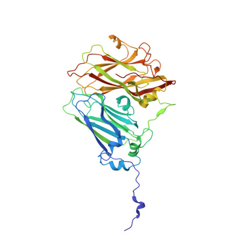
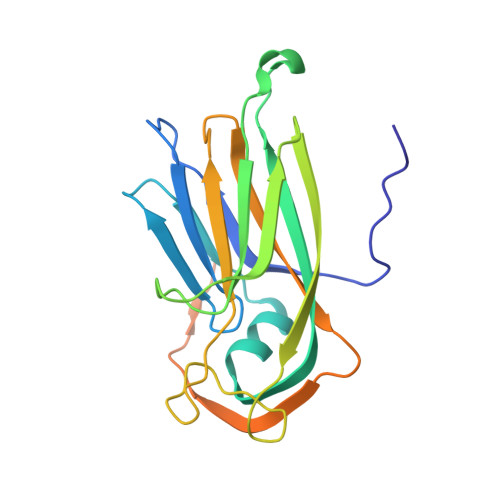
The cryo-electron microscopy structure of Broad Bean Stain Virus suggests a common capsid assembly mechanism among comoviruses.

Lecorre, F.Lai-Kee-Him, J.Blanc, S.Zeddam, J.L.Trapani, S.Bron, P.

(2019) Virology 530: 75-84

  • DOI: https://doi.org/10.1016/j.virol.2019.02.009
  • Primary Citation of Related Structures:  
    6QCC

  • PubMed Abstract: 

    The Broad bean stain virus (BBSV) is a member of the genus Comovirus infecting Fabaceae. The virus is transmitted through seed and by plant weevils causing severe and widespread disease worldwide. BBSV has a bipartite, positive-sense, single-stranded RNA genome encapsidated in icosahedral particles. We present here the cryo-electron microscopy reconstruction of the BBSV and an atomic model of the capsid proteins refined at 3.22 Å resolution. We identified residues involved in RNA/capsid interactions revealing a unique RNA genome organization. Inspection of the small coat protein C-terminal domain highlights a maturation cleavage between Leu567 and Leu568 and interactions of the C-terminal stretch with neighbouring small coat proteins within the capsid pentameric turrets. These interactions previously proposed to play a key role in the assembly of the Cowpea mosaic virus suggest a common capsid assembly mechanism throughout all comovirus species.


  • Organizational Affiliation

    Centre de Biochimie Structurale (CBS), INSERM, CNRS, Univ. Montpellier, 29 rue de Navacelles, 34090 Montpellier, France.


Macromolecules
Find similar proteins by:  (by identity cutoff)  |  3D Structure
Entity ID: 1
MoleculeChains Sequence LengthOrganismDetailsImage
Large coat-protein subunit377Broad bean stain virusMutation(s): 0 
UniProt
Find proteins for D0PSV7 (Broad bean stain virus)
Explore D0PSV7 
Go to UniProtKB:  D0PSV7
Entity Groups  
Sequence Clusters30% Identity50% Identity70% Identity90% Identity95% Identity100% Identity
UniProt GroupD0PSV7
Sequence Annotations
Expand
  • Reference Sequence
Find similar proteins by:  (by identity cutoff)  |  3D Structure
Entity ID: 2
MoleculeChains Sequence LengthOrganismDetailsImage
Small coat-protein subunit215Broad bean stain virusMutation(s): 0 
UniProt
Find proteins for D0PSV7 (Broad bean stain virus)
Explore D0PSV7 
Go to UniProtKB:  D0PSV7
Entity Groups  
Sequence Clusters30% Identity50% Identity70% Identity90% Identity95% Identity100% Identity
UniProt GroupD0PSV7
Sequence Annotations
Expand
  • Reference Sequence
Experimental Data & Validation

Experimental Data

  • Method: ELECTRON MICROSCOPY
  • Resolution: 3.22 Å
  • Aggregation State: PARTICLE 
  • Reconstruction Method: SINGLE PARTICLE 
EM Software:
TaskSoftware PackageVersion
RECONSTRUCTIONRELION3.0-beta-2
MODEL REFINEMENTPHENIX1.14-3260
MODEL REFINEMENTCoot0.8.9.1

Structure Validation

View Full Validation Report


Entry History & Funding Information

Deposition Data


Funding OrganizationLocationGrant Number
Government of New CaledoniaNew Caledonia--
French Infrastructure for Integrated Structural BiologyFranceANR-10-INBS-05
France BioImaging (FBI)FranceANR-10-INBS-04-01

Revision History  (Full details and data files)

  • Version 1.0: 2019-05-01
    Type: Initial release