6QBA | pdb_00006qba

Crystal Structure of Retinol-Binding Protein 4 (RBP4) in complex with non-retinoid ligand A1120 and engineered binding scaffold


Experimental Data Snapshot

  • Method: X-RAY DIFFRACTION
  • Resolution: 1.80 Å
  • R-Value Free: 
    0.221 (Depositor), 0.225 (DCC) 
  • R-Value Work: 
    0.188 (Depositor), 0.193 (DCC) 
  • R-Value Observed: 
    0.190 (Depositor) 

Starting Models: experimental
View more details

wwPDB Validation 3D Report Full Report

Validation slider image for 6QBA

Ligand Structure Quality Assessment 


This is version 1.3 of the entry. See complete history

Literature

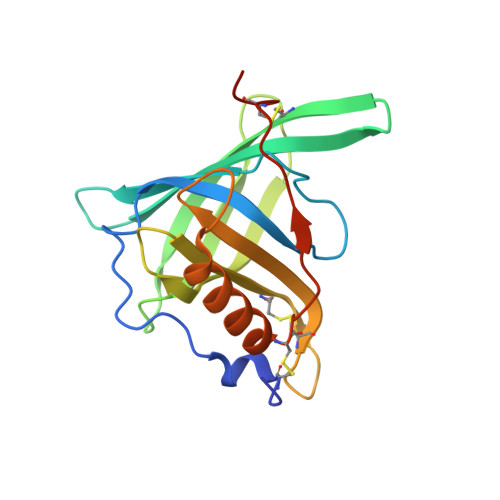
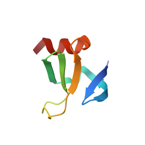
A conformation-specific ON-switch for controlling CAR T cells with an orally available drug.

Zajc, C.U.Dobersberger, M.Schaffner, I.Mlynek, G.Puhringer, D.Salzer, B.Djinovic-Carugo, K.Steinberger, P.De Sousa Linhares, A.Yang, N.J.Obinger, C.Holter, W.Traxlmayr, M.W.Lehner, M.

(2020) Proc Natl Acad Sci U S A 117: 14926-14935

  • DOI: https://doi.org/10.1073/pnas.1911154117
  • Primary Citation Related Structures: 
    6QBA

  • PubMed Abstract: 

    Molecular ON-switches in which a chemical compound induces protein-protein interactions can allow cellular function to be controlled with small molecules. ON-switches based on clinically applicable compounds and human proteins would greatly facilitate their therapeutic use. Here, we developed an ON-switch system in which the human retinol binding protein 4 (hRBP4) of the lipocalin family interacts with engineered hRBP4 binders in a small molecule-dependent manner. Two different protein scaffolds were engineered to bind to hRBP4 when loaded with the orally available small molecule A1120. The crystal structure of an assembled ON-switch shows that the engineered binder specifically recognizes the conformational changes induced by A1120 in two loop regions of hRBP4. We demonstrate that this conformation-specific ON-switch is highly dependent on the presence of A1120, as demonstrated by an ∼500-fold increase in affinity upon addition of the small molecule drug. Furthermore, the ON-switch successfully regulated the activity of primary human CAR T cells in vitro. We anticipate that lipocalin-based ON-switches have the potential to be broadly applied for the safe pharmacological control of cellular therapeutics.


  • Organizational Affiliation
    • St. Anna Children's Cancer Research Institute (CCRI), 1090 Vienna, Austria.

Macromolecule Content 

  • Total Structure Weight: 29.28 kDa 
  • Atom Count: 2,182 
  • Modeled Residue Count: 237 
  • Deposited Residue Count: 246 
  • Unique protein chains: 2

Macromolecules

Find similar proteins by:|  3D Structure
Entity ID: 1
MoleculeChains  Sequence LengthOrganismDetailsImage
Retinol-binding protein 4185Homo sapiensMutation(s): 0 
Gene Names: RBP4PRO2222
Membrane Entity: Yes 
UniProt & NIH Common Fund Data Resources
Find proteins for P02753 (Homo sapiens)
Explore P02753 
Go to UniProtKB:  P02753
PHAROS:  P02753
GTEx:  ENSG00000138207 
Entity Groups
Sequence Clusters30% Identity50% Identity70% Identity90% Identity95% Identity100% Identity
UniProt GroupP02753
Sequence Annotations
Expand
Reference Sequence
Find similar proteins by:|  3D Structure
Entity ID: 2
MoleculeChains  Sequence LengthOrganismDetailsImage
DNA-binding protein 7a61Saccharolobus solfataricusMutation(s): 0 
Gene Names: sso7a1sso7d-2SSO9180sso7a2sso7d-3SSO9535
EC: 3.1.27
UniProt
Find proteins for P61991 (Saccharolobus solfataricus (strain ATCC 35092 / DSM 1617 / JCM 11322 / P2))
Explore P61991 
Go to UniProtKB:  P61991
Entity Groups
Sequence Clusters30% Identity50% Identity70% Identity90% Identity95% Identity100% Identity
UniProt GroupP61991
Sequence Annotations
Expand
Reference Sequence

Small Molecules

Ligands 5 Unique
IDChains Name / Formula / InChI Key2D Diagram3D Interactions
2T1

Query on 2T1


Download:Ideal Coordinates CCD File
C [auth A]2-[({4-[2-(trifluoromethyl)phenyl]piperidin-1-yl}carbonyl)amino]benzoic acid
C20 H19 F3 N2 O3
MEAQCLPMSVEOQF-UHFFFAOYSA-N
PEG

Query on PEG


Download:Ideal Coordinates CCD File
I [auth A]DI(HYDROXYETHYL)ETHER
C4 H10 O3
MTHSVFCYNBDYFN-UHFFFAOYSA-N
IMD

Query on IMD


Download:Ideal Coordinates CCD File
D [auth A],
E [auth A],
F [auth A],
G [auth A],
H [auth A]
IMIDAZOLE
C3 H5 N2
RAXXELZNTBOGNW-UHFFFAOYSA-O
ZN

Query on ZN


Download:Ideal Coordinates CCD File
J [auth A],
K [auth A],
L [auth A]
ZINC ION
Zn
PTFCDOFLOPIGGS-UHFFFAOYSA-N
ACT

Query on ACT


Download:Ideal Coordinates CCD File
M [auth B]ACETATE ION
C2 H3 O2
QTBSBXVTEAMEQO-UHFFFAOYSA-M
Binding Affinity Annotations 
IDSourceBinding Affinity
2T1 BindingDB:  6QBA IC50: min: 0.16, max: 122 (nM) from 5 assay(s)

Experimental Data & Validation

Experimental Data

  • Method: X-RAY DIFFRACTION
  • Resolution: 1.80 Å
  • R-Value Free:  0.221 (Depositor), 0.225 (DCC) 
  • R-Value Work:  0.188 (Depositor), 0.193 (DCC) 
  • R-Value Observed: 0.190 (Depositor) 
Space Group: P 21 21 21
Unit Cell:
Length ( Å )Angle ( ˚ )
a = 44.456α = 90
b = 77.614β = 90
c = 80.509γ = 90
Software Package:
Software NamePurpose
PHENIXrefinement
XDSdata reduction
PHASERphasing
EDNAdata collection
XDSdata scaling

Structure Validation

View Full Validation Report


Ligand Structure Quality Assessment 


Entry History 

Deposition Data

Revision History  (Full details and data files)

  • Version 1.0: 2020-06-03
    Type: Initial release
  • Version 1.1: 2020-12-16
    Changes: Database references, Derived calculations
  • Version 1.2: 2024-01-24
    Changes: Data collection, Database references, Refinement description
  • Version 1.3: 2024-11-06
    Changes: Structure summary