6Q53

CRYSTAL STRUCTURE OF THE LIGHT-HARVESTING COMPLEX II (B800-850) FROM Ectothiorhodospira haloalkaliphila


Experimental Data Snapshot

  • Method: X-RAY DIFFRACTION
  • Resolution: 3.70 Å
  • R-Value Free: 0.277 
  • R-Value Work: 0.240 
  • R-Value Observed: 0.242 

wwPDB Validation   3D Report Full Report


Ligand Structure Quality Assessment 


This is version 1.1 of the entry. See complete history


Literature

Controlling Photosynthetic Excitons by Selective Pigment Photooxidation.

Leiger, K.Linnanto, J.M.Ratsep, M.Timpmann, K.Ashikhmin, A.A.Moskalenko, A.A.Fufina, T.Y.Gabdulkhakov, A.G.Freiberg, A.

(2019) J Phys Chem B 123: 29-38

  • DOI: https://doi.org/10.1021/acs.jpcb.8b08083
  • Primary Citation of Related Structures:  
    6Q53

  • PubMed Abstract: 

    As a basis of photosynthesis, photoinduced oxidation of (bacterio)chlorophyll molecules in the special reaction center complexes has been a subject of extensive research. In contrast, the generally harmful photooxidation of antenna chromoproteins has received much less attention. Here, we have established the permanent structural changes in the LH2 antenna bacteriochlorophyll-protein complex from a sulfur photosynthetic purple bacterium Ectothiorhodospira haloalkaliphila taking place at physiological conditions upon intense optical irradiation. To this end, a crystal structure of the LH2 complex from E. haloalkaliphila was first resolved by X-ray diffraction to 3.7 Å, verifying a great similarity with the earlier structure from Phaesporillum molischianum. Analysis of the various steady-state and picosecond time-resolved optical spectroscopy data and related model simulations then confirmed that the major spectral effects observed-bleaching and blue-shifting of the B850 exciton band and correlated emergence of a higher-energy C700 exciton band-are associated with photooxidation of increasing numbers of B850 bacteriochlorophylls into 3-acetyl-chlorophylls, with no noticeable damage to the pigment-binding protein scaffold. A prospective noninvasive method for an in situ optical control of excitons by selective photooxidation of pigment chromophores was thus revealed and demonstrated in a structurally well-defined native system.


  • Organizational Affiliation

    Institute of Physics , University of Tartu , W. Ostwaldi 1 , Tartu 50411 , Estonia.


Macromolecules
Find similar proteins by:  (by identity cutoff)  |  3D Structure
Entity ID: 1
MoleculeChains Sequence LengthOrganismDetailsImage
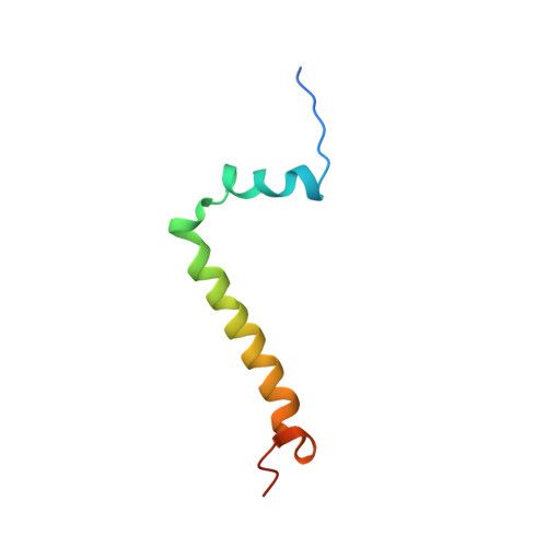
light-harvesting protein subunit alphaA,
C [auth D]
61Ectothiorhodospira haloalkaliphilaMutation(s): 0 
Entity Groups  
Sequence Clusters30% Identity50% Identity70% Identity90% Identity95% Identity100% Identity
Sequence Annotations
Expand
  • Reference Sequence
Find similar proteins by:  (by identity cutoff)  |  3D Structure
Entity ID: 2
MoleculeChains Sequence LengthOrganismDetailsImage
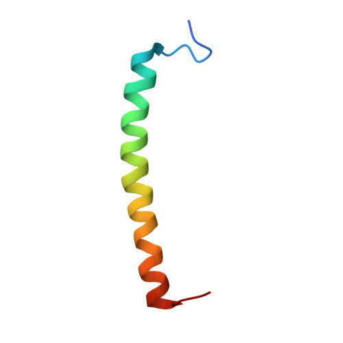
Light-harvesting protein B:800-850 subunit betaB,
D [auth E]
45Halorhodospira halochloris str. AMutation(s): 0 
Gene Names: M911_15650
Membrane Entity: Yes 
UniProt
Find proteins for W8L932 (Ectothiorhodospira haloalkaliphila)
Explore W8L932 
Go to UniProtKB:  W8L932
Entity Groups  
Sequence Clusters30% Identity50% Identity70% Identity90% Identity95% Identity100% Identity
UniProt GroupW8L932
Sequence Annotations
Expand
  • Reference Sequence
Small Molecules
Ligands 3 Unique
IDChains Name / Formula / InChI Key2D Diagram3D Interactions
BCL
Query on BCL

Download Ideal Coordinates CCD File 
E [auth A]
F [auth A]
H [auth B]
J [auth D]
K [auth D]
E [auth A],
F [auth A],
H [auth B],
J [auth D],
K [auth D],
M [auth E]
BACTERIOCHLOROPHYLL A
C55 H74 Mg N4 O6
DSJXIQQMORJERS-AGGZHOMASA-M
LYC
Query on LYC

Download Ideal Coordinates CCD File 
G [auth A],
L [auth E]
LYCOPENE
C40 H56
OAIJSZIZWZSQBC-GYZMGTAESA-N
DET
Query on DET

Download Ideal Coordinates CCD File 
I [auth B],
N [auth E],
O [auth E]
UNDECYLAMINE-N,N-DIMETHYL-N-OXIDE
C13 H29 N O
OZHBUVQCJMARBN-UHFFFAOYSA-N
Experimental Data & Validation

Experimental Data

  • Method: X-RAY DIFFRACTION
  • Resolution: 3.70 Å
  • R-Value Free: 0.277 
  • R-Value Work: 0.240 
  • R-Value Observed: 0.242 
  • Space Group: P 4 3 2
Unit Cell:
Length ( Å )Angle ( ˚ )
a = 177.24α = 90
b = 177.24β = 90
c = 177.24γ = 90
Software Package:
Software NamePurpose
PHENIXrefinement
XDSdata reduction
XSCALEdata scaling
PHASERphasing

Structure Validation

View Full Validation Report


Ligand Structure Quality Assessment 


Entry History & Funding Information

Deposition Data


Funding OrganizationLocationGrant Number
Russian Federationprogram of the Presidium of the Russian Academy of Sciences Molecular and Cell Biology and Postgenomic Technologies

Revision History  (Full details and data files)

  • Version 1.0: 2019-10-16
    Type: Initial release
  • Version 1.1: 2024-01-24
    Changes: Data collection, Database references, Refinement description