6Q3R | pdb_00006q3r

ASPERGILLUS ACULEATUS GALACTANASE


Experimental Data Snapshot

  • Method: X-RAY DIFFRACTION
  • Resolution: 2.69 Å
  • R-Value Free: 
    0.211 (Depositor), 0.214 (DCC) 
  • R-Value Work: 
    0.145 (Depositor), 0.154 (DCC) 
  • R-Value Observed: 
    0.148 (Depositor) 

Starting Model: experimental
View more details

wwPDB Validation 3D Report Full Report

Validation slider image for 6Q3R

This is version 2.2 of the entry. See complete history

Literature

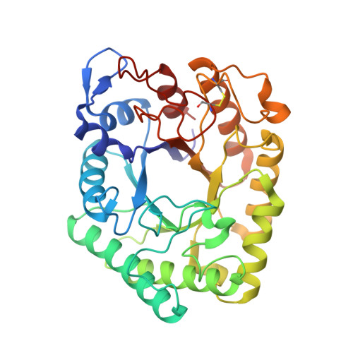
Structure of Aspergillus aculeatus beta-1,4-galactanase in complex with galactobiose.

Torpenholt, S.Poulsen, J.C.N.Muderspach, S.J.De Maria, L.Lo Leggio, L.

(2019) Acta Crystallogr F Struct Biol Commun 75: 399-404

  • DOI: https://doi.org/10.1107/S2053230X19005612
  • Primary Citation Related Structures: 
    6Q3R

  • PubMed Abstract: 

    β-1,4-Galactanases are glycoside hydrolases that are involved in the degradation of pectin and belong to family 53 in the classification of glycoside hydrolases. Previous studies have elucidated the structures of several fungal and two bacterial galactanases, while biochemical studies have indicated differences in the product profiles of different members of the family. Structural studies of ligand complexes have to date been limited to the bacterial members of the family. Here, the first structure of a fungal galactanase in complex with a disaccharide is presented. Galactobiose binds to subsites -1 and -2, thus improving our understanding of ligand binding to galactanases.


  • Organizational Affiliation
    • Department of Chemistry, University of Copenhagen, Universitetsparken 5, 2100 Copenhagen, Denmark.

Macromolecule Content 

  • Total Structure Weight: 77.9 kDa 
  • Atom Count: 5,711 
  • Modeled Residue Count: 668 
  • Deposited Residue Count: 668 
  • Unique protein chains: 1

Macromolecules

Find similar proteins by:|  3D Structure
Entity ID: 1
MoleculeChains  Sequence LengthOrganismDetailsImage
Arabinogalactan endo-beta-1,4-galactanase
A, B
334Aspergillus aculeatusMutation(s): 0 
EC: 3.2.1.89
UniProt
Find proteins for P48842 (Aspergillus aculeatus)
Explore P48842 
Go to UniProtKB:  P48842
Entity Groups
Sequence Clusters30% Identity50% Identity70% Identity90% Identity95% Identity100% Identity
UniProt GroupP48842
Glycosylation
Glycosylation Sites: 1
Sequence Annotations
Expand
Reference Sequence

Oligosaccharides

Help  
Entity ID: 2
MoleculeChains Length2D Diagram GlycosylationD Interactions
beta-D-mannopyranose-(1-4)-2-acetamido-2-deoxy-beta-D-glucopyranose-(1-4)-2-acetamido-2-deoxy-beta-D-glucopyranose
C, F
3N-Glycosylation
Glycosylation Resources
GlyTouCan: G15407YE
GlyCosmos: G15407YE
GlyGen: G15407YE
Entity ID: 3
MoleculeChains Length2D Diagram GlycosylationD Interactions
beta-D-galactopyranose-(1-4)-beta-D-galactopyranose
D, E
2N/A
Glycosylation Resources
GlyTouCan: G08806VI
GlyCosmos: G08806VI
GlyGen: G08806VI

Small Molecules

Ligands 7 Unique
IDChains Name / Formula / InChI Key2D Diagram3D Interactions
PG6

Query on PG6


Download:Ideal Coordinates CCD File
W [auth A]1-(2-METHOXY-ETHOXY)-2-{2-[2-(2-METHOXY-ETHOXY]-ETHOXY}-ETHANE
C12 H26 O6
DMDPGPKXQDIQQG-UHFFFAOYSA-N
PG4

Query on PG4


Download:Ideal Coordinates CCD File
CA [auth B],
M [auth A]
TETRAETHYLENE GLYCOL
C8 H18 O5
UWHCKJMYHZGTIT-UHFFFAOYSA-N
PGE

Query on PGE


Download:Ideal Coordinates CCD File
KA [auth B]TRIETHYLENE GLYCOL
C6 H14 O4
ZIBGPFATKBEMQZ-UHFFFAOYSA-N
PEG

Query on PEG


Download:Ideal Coordinates CCD File
G [auth A],
H [auth A],
X [auth B],
Y [auth B]
DI(HYDROXYETHYL)ETHER
C4 H10 O3
MTHSVFCYNBDYFN-UHFFFAOYSA-N
EDO

Query on EDO


Download:Ideal Coordinates CCD File
DA [auth B]
EA [auth B]
FA [auth B]
GA [auth B]
HA [auth B]
DA [auth B],
EA [auth B],
FA [auth B],
GA [auth B],
HA [auth B],
IA [auth B],
N [auth A],
O [auth A],
P [auth A],
Q [auth A],
R [auth A],
S [auth A],
T [auth A]
1,2-ETHANEDIOL
C2 H6 O2
LYCAIKOWRPUZTN-UHFFFAOYSA-N
ACT

Query on ACT


Download:Ideal Coordinates CCD File
JA [auth B],
U [auth A],
V [auth A]
ACETATE ION
C2 H3 O2
QTBSBXVTEAMEQO-UHFFFAOYSA-M
CA

Query on CA


Download:Ideal Coordinates CCD File
AA [auth B]
BA [auth B]
I [auth A]
J [auth A]
K [auth A]
AA [auth B],
BA [auth B],
I [auth A],
J [auth A],
K [auth A],
L [auth A],
Z [auth B]
CALCIUM ION
Ca
BHPQYMZQTOCNFJ-UHFFFAOYSA-N

Biologically Interesting Molecules (External Reference) 

1 Unique

Experimental Data & Validation

Experimental Data

  • Method: X-RAY DIFFRACTION
  • Resolution: 2.69 Å
  • R-Value Free:  0.211 (Depositor), 0.214 (DCC) 
  • R-Value Work:  0.145 (Depositor), 0.154 (DCC) 
  • R-Value Observed: 0.148 (Depositor) 
Space Group: I 41 2 2
Unit Cell:
Length ( Å )Angle ( ˚ )
a = 200.2α = 90
b = 200.2β = 90
c = 106.83γ = 90
Software Package:
Software NamePurpose
REFMACrefinement
XDSdata reduction
XSCALEdata scaling
PHASERphasing

Structure Validation

View Full Validation Report


Entry History 

Deposition Data

Revision History  (Full details and data files)

  • Version 1.0: 2019-06-12
    Type: Initial release
  • Version 1.1: 2019-06-26
    Changes: Data collection, Database references
  • Version 2.0: 2020-07-29
    Type: Remediation
    Reason: Carbohydrate remediation
    Changes: Advisory, Atomic model, Data collection, Derived calculations, Structure summary
  • Version 2.1: 2024-01-24
    Changes: Data collection, Database references, Refinement description, Structure summary
  • Version 2.2: 2024-11-06
    Changes: Structure summary