6Q2D

Crystal structure of Methanobrevibacter smithii Dph2 in complex with Methanobrevibacter smithii elongation factor 2


Experimental Data Snapshot

  • Method: X-RAY DIFFRACTION
  • Resolution: 3.45 Å
  • R-Value Free: 0.267 
  • R-Value Work: 0.224 
  • R-Value Observed: 0.226 

wwPDB Validation   3D Report Full Report


This is version 1.4 of the entry. See complete history


Literature

The Crystal Structure of Dph2 in Complex with Elongation Factor 2 Reveals the Structural Basis for the First Step of Diphthamide Biosynthesis.

Fenwick, M.K.Dong, M.Lin, H.Ealick, S.E.

(2019) Biochemistry 58: 4343-4351

  • DOI: https://doi.org/10.1021/acs.biochem.9b00718
  • Primary Citation of Related Structures:  
    6Q2D, 6Q2E

  • PubMed Abstract: 

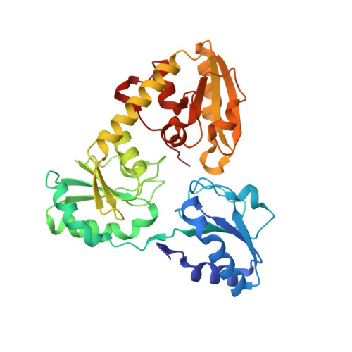
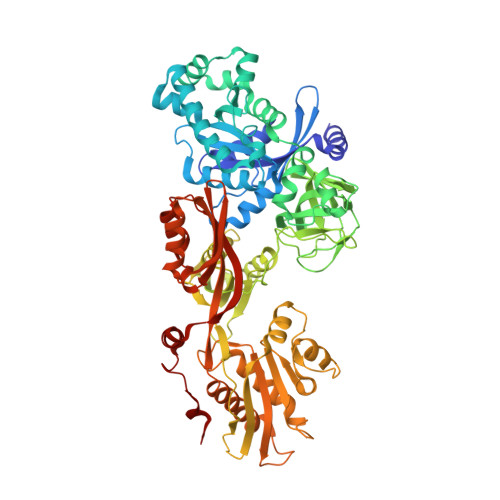
    Elongation factor 2 (EF-2), a five-domain, GTP-dependent ribosomal translocase of archaebacteria and eukaryotes, undergoes post-translational modification to form diphthamide on a specific histidine residue in domain IV prior to binding the ribosome. The first step of diphthamide biosynthesis in archaebacteria is catalyzed by Dph2, a homodimeric radical S -adenosylmethionine (SAM) enzyme having a noncanonical architecture. Here, we describe a 3.5 Å resolution crystal structure of the Methanobrevibacter smithii ( Ms ) Dph2 homodimer bound to two molecules of Ms EF-2, one of which is ordered and the other largely disordered. Ms EF-2 is bound to both protomers of Ms Dph2, with domain IV bound to the active site of one protomer and domain III bound to a surface α-helix of an adjacent protomer. The histidine substrate of domain IV is inserted into the active site, which reveals for the first time the architecture of the Dph2 active site in complex with its target substrate. We also determined a high-resolution crystal structure of isolated Ms Dph2 bound to 5'-methylthioadenosine that shows a conserved arginine residue preoriented by conserved phenylalanine and aspartate residues for binding the carboxylate group of SAM. Mutagenesis experiments suggest that the arginine plays an important role in the first step of diphthamide biosynthesis.


Macromolecules
Find similar proteins by:  (by identity cutoff)  |  3D Structure
Entity ID: 1
MoleculeChains Sequence LengthOrganismDetailsImage
2-(3-amino-3-carboxypropyl)histidine synthase
A, B
337Methanobrevibacter smithiiMutation(s): 0 
Gene Names: Msm_1358
EC: 2.5.1.108
UniProt
Find proteins for A5UMY5 (Methanobrevibacter smithii (strain ATCC 35061 / DSM 861 / OCM 144 / PS))
Explore A5UMY5 
Go to UniProtKB:  A5UMY5
Entity Groups  
Sequence Clusters30% Identity50% Identity70% Identity90% Identity95% Identity100% Identity
UniProt GroupA5UMY5
Sequence Annotations
Expand
  • Reference Sequence
Find similar proteins by:  (by identity cutoff)  |  3D Structure
Entity ID: 2
MoleculeChains Sequence LengthOrganismDetailsImage
Elongation factor 2C,
D [auth F]
733Methanobrevibacter smithiiMutation(s): 0 
Gene Names: fusABK798_06620
UniProt
Find proteins for A0A2H4U7K7 (Methanobrevibacter smithii)
Explore A0A2H4U7K7 
Go to UniProtKB:  A0A2H4U7K7
Entity Groups  
Sequence Clusters30% Identity50% Identity70% Identity90% Identity95% Identity100% Identity
UniProt GroupA0A2H4U7K7
Sequence Annotations
Expand
  • Reference Sequence
Experimental Data & Validation

Experimental Data

  • Method: X-RAY DIFFRACTION
  • Resolution: 3.45 Å
  • R-Value Free: 0.267 
  • R-Value Work: 0.224 
  • R-Value Observed: 0.226 
  • Space Group: C 2 2 21
Unit Cell:
Length ( Å )Angle ( ˚ )
a = 100.899α = 90
b = 322.979β = 90
c = 172.432γ = 90
Software Package:
Software NamePurpose
PHENIXrefinement
PDB_EXTRACTdata extraction
XDSdata reduction
Aimlessdata scaling
PHASERphasing

Structure Validation

View Full Validation Report


Entry History & Funding Information

Deposition Data


Funding OrganizationLocationGrant Number
Department of Energy (DOE, United States)United StatesDE-AC02-06CH11357
National Institutes of Health/National Institute of General Medical Sciences (NIH/NIGMS)United StatesP30GM124165

Revision History  (Full details and data files)

  • Version 1.0: 2019-10-16
    Type: Initial release
  • Version 1.1: 2019-11-06
    Changes: Data collection, Database references
  • Version 1.2: 2019-11-13
    Changes: Data collection, Database references
  • Version 1.3: 2019-12-04
    Changes: Author supporting evidence
  • Version 1.4: 2023-10-11
    Changes: Data collection, Database references, Refinement description