


Structure of truncated hemolysin A variant F80L
Weaver, T.M., Novak, W.R.P., Bhattacharyya, B., Woods, C.N., Grilley, D.P., Wimmer, M.R.To be published.
Experimental Data Snapshot
Starting Model: experimental
View more details
wwPDB Validation 3D Report Full Report

Entity ID: 1 | |||||
|---|---|---|---|---|---|
| Molecule | Chains | Sequence Length | Organism | Details | Image |
| Hemolysin | 242 | Proteus mirabilis | Mutation(s): 1 Gene Names: hpmA | 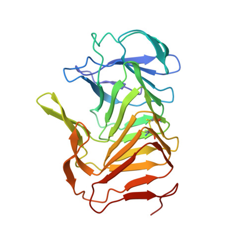 | |
UniProt | |||||
Entity Groups | |||||
| Sequence Clusters | 30% Identity50% Identity70% Identity90% Identity95% Identity100% Identity | ||||
| UniProt Group | P16466 | ||||
Sequence AnnotationsExpand | |||||
Reference Sequence | |||||
| Length ( Å ) | Angle ( ˚ ) |
|---|---|
| a = 51.55 | α = 90 |
| b = 34.011 | β = 101.02 |
| c = 59.48 | γ = 90 |
| Software Name | Purpose |
|---|---|
| PHENIX | refinement |
| PDB_EXTRACT | data extraction |
| HKL-2000 | data reduction |
| HKL-2000 | data scaling |
| PHENIX | phasing |
| Coot | model building |
| Funding Organization | Location | Grant Number |
|---|---|---|
| National Science Foundation (NSF, United States) | United States | MCB1050435 |
| National Science Foundation (NSF, United States) | United States | REU1434473 |


