6PSY | pdb_00006psy

Cryo-EM structure of S. cerevisiae Drs2p-Cdc50p in the autoinhibited apo form


Experimental Data Snapshot

  • Method: ELECTRON MICROSCOPY
  • Resolution: 2.80 Å
  • Aggregation State: PARTICLE 
  • Reconstruction Method: SINGLE PARTICLE 

wwPDB Validation 3D Report Full Report

Validation slider image for 6PSY

This is version 2.1 of the entry. See complete history

Literature

Autoinhibition and activation mechanisms of the eukaryotic lipid flippase Drs2p-Cdc50p.

Bai, L.Kovach, A.You, Q.Hsu, H.C.Zhao, G.Li, H.

(2019) Nat Commun 10: 4142-4142

  • DOI: https://doi.org/10.1038/s41467-019-12191-9
  • Primary Citation Related Structures: 
    6PSX, 6PSY

  • PubMed Abstract: 

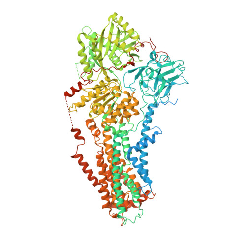
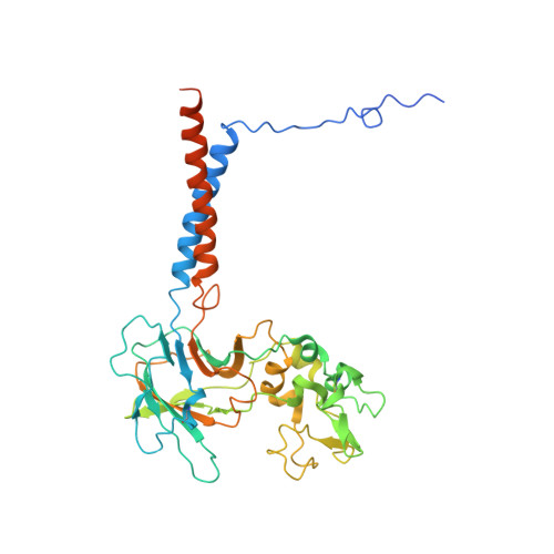
    The heterodimeric eukaryotic Drs2p-Cdc50p complex is a lipid flippase that maintains cell membrane asymmetry. The enzyme complex exists in an autoinhibited form in the absence of an activator and is specifically activated by phosphatidylinositol-4-phosphate (PI4P), although the underlying mechanisms have been unclear. Here we report the cryo-EM structures of intact Drs2p-Cdc50p isolated from S. cerevisiae in apo form and in the PI4P-activated form at 2.8 Å and 3.3 Å resolution, respectively. The structures reveal that the Drs2p C-terminus lines a long groove in the cytosolic regulatory region to inhibit the flippase activity. PIP4 binding in a cytosol-proximal membrane region triggers a 90° rotation of a cytosolic helix switch that is located just upstream of the inhibitory C-terminal peptide. The rotation of the helix switch dislodges the C-terminus from the regulatory region, activating the flippase.


  • Organizational Affiliation
    • Structural Biology Program, Van Andel Research Institute, Grand Rapids, MI, 49503, USA. Lin.Bai@vai.org.

Macromolecule Content 

  • Total Structure Weight: 199.83 kDa 
  • Atom Count: 11,540 
  • Modeled Residue Count: 1,429 
  • Deposited Residue Count: 1,746 
  • Unique protein chains: 2

Macromolecules

Find similar proteins by:|  3D Structure
Entity ID: 1
MoleculeChains  Sequence LengthOrganismDetailsImage
Probable phospholipid-transporting ATPase DRS21,355Saccharomyces cerevisiae W303Mutation(s): 0 
Gene Names: DRS2YAL026CFUN38
EC: 7.6.2.1
Membrane Entity: Yes 
UniProt
Find proteins for P39524 (Saccharomyces cerevisiae (strain ATCC 204508 / S288c))
Explore P39524 
Go to UniProtKB:  P39524
Entity Groups
Sequence Clusters30% Identity50% Identity70% Identity90% Identity95% Identity100% Identity
UniProt GroupP39524
Sequence Annotations
Expand
Reference Sequence
Find similar proteins by:|  3D Structure
Entity ID: 2
MoleculeChains  Sequence LengthOrganismDetailsImage
Cell division control protein 50B [auth E]391Saccharomyces cerevisiae W303Mutation(s): 0 
Gene Names: CDC50YCR094WYCR94W
Membrane Entity: Yes 
UniProt
Find proteins for P25656 (Saccharomyces cerevisiae (strain ATCC 204508 / S288c))
Explore P25656 
Go to UniProtKB:  P25656
Entity Groups
Sequence Clusters30% Identity50% Identity70% Identity90% Identity95% Identity100% Identity
UniProt GroupP25656
Glycosylation
Glycosylation Sites: 3Go to GlyGen: P25656-1
Sequence Annotations
Expand
Reference Sequence

Oligosaccharides

Help  
Entity ID: 3
MoleculeChains Length2D Diagram GlycosylationD Interactions
2-acetamido-2-deoxy-beta-D-glucopyranose-(1-4)-2-acetamido-2-deoxy-beta-D-glucopyranoseC [auth B]2N-Glycosylation
Glycosylation Resources
GlyTouCan: G42666HT
GlyCosmos: G42666HT
GlyGen: G42666HT

Small Molecules

Experimental Data & Validation

Experimental Data

  • Method: ELECTRON MICROSCOPY
  • Resolution: 2.80 Å
  • Aggregation State: PARTICLE 
  • Reconstruction Method: SINGLE PARTICLE 
EM Software:
TaskSoftware PackageVersion
MODEL REFINEMENTPHENIX
MODEL REFINEMENTCoot
RECONSTRUCTIONCoot
RECONSTRUCTIONUCSF Chimera

Structure Validation

View Full Validation Report


Entry History 

& Funding Information

Deposition Data

  • Released Date: 2019-09-25 
  • Deposition Author(s): Bai, L., Li, H.

Funding OrganizationLocationGrant Number
National Institutes of Health/National Cancer Institute (NIH/NCI)United StatesR01-CA231466

Revision History  (Full details and data files)

  • Version 1.0: 2019-09-25
    Type: Initial release
  • Version 1.1: 2019-12-04
    Changes: Author supporting evidence
  • Version 2.0: 2020-07-29
    Type: Remediation
    Reason: Carbohydrate remediation
    Changes: Atomic model, Data collection, Derived calculations, Structure summary
  • Version 2.1: 2024-10-23
    Changes: Data collection, Database references, Structure summary