6PON | pdb_00006pon

CRYSTAL STRUCTURE OF THE N-TERMINAL DOMAIN OF FIBRONECTIN- BINDING PROTEIN PAVA FROM STREPTOCOCCUS PNEUMONIAE


Experimental Data Snapshot

  • Method: X-RAY DIFFRACTION
  • Resolution: 2.40 Å
  • R-Value Free: 
    0.237 (Depositor) 
  • R-Value Work: 
    0.167 (Depositor) 
  • R-Value Observed: 
    0.170 (Depositor) 

Starting Model: experimental
View more details

wwPDB Validation   3D Report Full Report

Currently 6PON does not have a validation slider image.


This is version 1.4 of the entry. See complete history


Literature

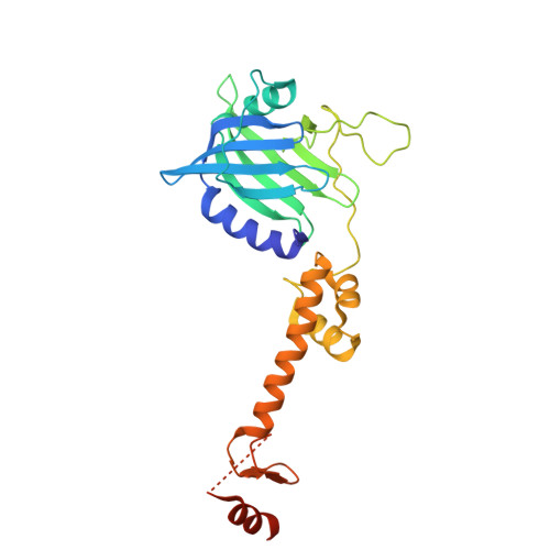
Crystal structure of the N-terminal domain of the fibronectin-binding protein PavA from Streptococcus pneumoniae.

Manne, K.Narayana, S.V.L.Chattopadhyay, D.

(2019) Acta Crystallogr F Struct Biol Commun 75: 657-662

  • DOI: https://doi.org/10.1107/S2053230X19012160
  • Primary Citation of Related Structures:  
    6PON

  • PubMed Abstract: 

    The Gram-positive bacterium Streptococcus pneumoniae, a major human pathogen, is a regular colonizer of the upper and lower respiratory tracts. Pneumococcal adherence and virulence factor A (PavA), a fibronectin-binding bacterial protein, from S. pneumoniae is an important facilitator of its colonization of host cells. In this study, the crystal structure of the N-terminal domain of PavA (SpPavA-N) determined at a resolution of 2.39 Å is reported. Each monomer of the dimeric protein consists of two domains (domains I and II) and a short α-helix (α6) at the C-terminus that are connected by elongated loops. Comparison of the SpPavA-N structure with that of its homolog from Streptococcus suis (FBPS-N) revealed differences in α5, α6 and the domain II/α6 inter-loop region within domain II. The α5 helix of FBPS-N folds back toward domain I, whereas in SpPavA-N it adopts an elongated rod shape.


  • Organizational Affiliation
    • Center for Biophysical Sciences and Engineering, University of Alabama at Birmingham, Birmingham, AL 35294, USA.

Macromolecules
Find similar proteins by:  (by identity cutoff)  |  3D Structure
Entity ID: 1
MoleculeChains Sequence LengthOrganismDetailsImage
Adherence and virulence protein A
A, B
274Streptococcus pneumoniaeMutation(s): 0 
Gene Names: pavA
UniProt
Find proteins for Q8DQ36 (Streptococcus pneumoniae (strain ATCC BAA-255 / R6))
Explore Q8DQ36 
Go to UniProtKB:  Q8DQ36
Entity Groups  
Sequence Clusters30% Identity50% Identity70% Identity90% Identity95% Identity100% Identity
UniProt GroupQ8DQ36
Sequence Annotations
Expand
  • Reference Sequence
Experimental Data & Validation

Experimental Data

  • Method: X-RAY DIFFRACTION
  • Resolution: 2.40 Å
  • R-Value Free:  0.237 (Depositor) 
  • R-Value Work:  0.167 (Depositor) 
  • R-Value Observed: 0.170 (Depositor) 
Space Group: P 1 21 1
Unit Cell:
Length ( Å )Angle ( ˚ )
a = 75.873α = 90
b = 46.814β = 90.931
c = 83.678γ = 90
Software Package:
Software NamePurpose
PHENIXrefinement
PHENIXrefinement
HKL-3000data reduction
HKL-3000data scaling
PHENIXphasing

Structure Validation

View Full Validation Report

Currently 6PON does not have a validation slider image.


Entry History & Funding Information

Deposition Data


Funding OrganizationLocationGrant Number
National Institutes of Health/National Institute Of Allergy and Infectious Diseases (NIH/NIAID)United StatesR01- AI106808

Revision History  (Full details and data files)

  • Version 1.0: 2019-07-31
    Type: Initial release
  • Version 1.1: 2019-09-04
    Changes: Data collection, Database references, Structure summary
  • Version 1.2: 2019-10-16
    Changes: Data collection, Database references
  • Version 1.3: 2019-12-18
    Changes: Author supporting evidence
  • Version 1.4: 2023-10-11
    Changes: Data collection, Database references, Refinement description