6PKF

Myocilin OLF mutant N428E/D478K


Experimental Data Snapshot

  • Method: X-RAY DIFFRACTION
  • Resolution: 1.48 Å
  • R-Value Free: 0.181 
  • R-Value Work: 0.150 
  • R-Value Observed: 0.152 

wwPDB Validation   3D Report Full Report

Currently 6PKF does not have a validation slider image.


This is version 1.3 of the entry. See complete history


Literature

Calcium-ligand variants of the myocilin olfactomedin propeller selected from invertebrate phyla reveal cross-talk with N-terminal blade and surface helices.

Hill, S.E.Cho, H.Raut, P.Lieberman, R.L.

(2019) Acta Crystallogr D Struct Biol 75: 817-824

  • DOI: https://doi.org/10.1107/S205979831901074X
  • Primary Citation of Related Structures:  
    6PKD, 6PKE, 6PKF

  • PubMed Abstract: 

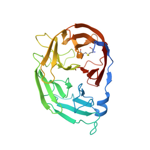
    Olfactomedins are a family of modular proteins found in multicellular organisms that all contain five-bladed β-propeller olfactomedin (OLF) domains. In support of differential functions for the OLF propeller, the available crystal structures reveal that only some OLF domains harbor an internal calcium-binding site with ligands derived from a triad of residues. For the myocilin OLF domain (myoc-OLF), ablation of the ion-binding site (triad Asp, Asn, Asp) by altering the coordinating residues affects the stability and overall structure, in one case leading to misfolding and glaucoma. Bioinformatics analysis reveals a variety of triads with possible ion-binding characteristics lurking in OLF domains in invertebrate chordates such as Arthropoda (Asp-Glu-Ser), Nematoda (Asp-Asp-His) and Echinodermata (Asp-Glu-Lys). To test ion binding and to extend the observed connection between ion binding and distal structural rearrangements, consensus triads from these phyla were installed in the myoc-OLF. All three protein variants exhibit wild-type-like or better stability, but their calcium-binding properties differ, concomitant with new structural deviations from wild-type myoc-OLF. Taken together, the results indicate that calcium binding is not intrinsically destabilizing to myoc-OLF or required to observe a well ordered side helix, and that ion binding is a differential feature that may underlie the largely elusive biological function of OLF propellers.


  • Organizational Affiliation

    School of Chemistry and Biochemistry, Georgia Institute of Technology, 901 Atlantic Drive NW, Atlanta, GA 30332-0400, USA.


Macromolecules
Find similar proteins by:  (by identity cutoff)  |  3D Structure
Entity ID: 1
MoleculeChains Sequence LengthOrganismDetailsImage
Myocilin277Homo sapiensMutation(s): 2 
Gene Names: MYOCGLC1ATIGR
UniProt & NIH Common Fund Data Resources
Find proteins for Q99972 (Homo sapiens)
Explore Q99972 
Go to UniProtKB:  Q99972
PHAROS:  Q99972
GTEx:  ENSG00000034971 
Entity Groups  
Sequence Clusters30% Identity50% Identity70% Identity90% Identity95% Identity100% Identity
UniProt GroupQ99972
Sequence Annotations
Expand
  • Reference Sequence
Small Molecules
Ligands 1 Unique
IDChains Name / Formula / InChI Key2D Diagram3D Interactions
GOL
Query on GOL

Download Ideal Coordinates CCD File 
B [auth A]GLYCEROL
C3 H8 O3
PEDCQBHIVMGVHV-UHFFFAOYSA-N
Experimental Data & Validation

Experimental Data

  • Method: X-RAY DIFFRACTION
  • Resolution: 1.48 Å
  • R-Value Free: 0.181 
  • R-Value Work: 0.150 
  • R-Value Observed: 0.152 
  • Space Group: P 21 21 21
Unit Cell:
Length ( Å )Angle ( ˚ )
a = 45.506α = 90
b = 58.688β = 90
c = 89.505γ = 90
Software Package:
Software NamePurpose
HKL-2000data reduction
HKL-2000data scaling
PHENIXrefinement
PDB_EXTRACTdata extraction
PHASERphasing

Structure Validation

View Full Validation Report

Currently 6PKF does not have a validation slider image.


Entry History & Funding Information

Deposition Data


Funding OrganizationLocationGrant Number
National Institutes of Health/National Eye Institute (NIH/NEI)United StatesR01EY021205

Revision History  (Full details and data files)

  • Version 1.0: 2019-09-11
    Type: Initial release
  • Version 1.1: 2019-11-13
    Changes: Database references
  • Version 1.2: 2019-12-11
    Changes: Author supporting evidence
  • Version 1.3: 2023-10-11
    Changes: Data collection, Database references, Refinement description