6PJQ

Time-resolved structural snapshot of proteolysis by GlpG inside the membrane


Experimental Data Snapshot

  • Method: X-RAY DIFFRACTION
  • Resolution: 2.50 Å
  • R-Value Free: 0.288 
  • R-Value Work: 0.241 
  • R-Value Observed: 0.243 

wwPDB Validation   3D Report Full Report


This is version 1.2 of the entry. See complete history


Literature

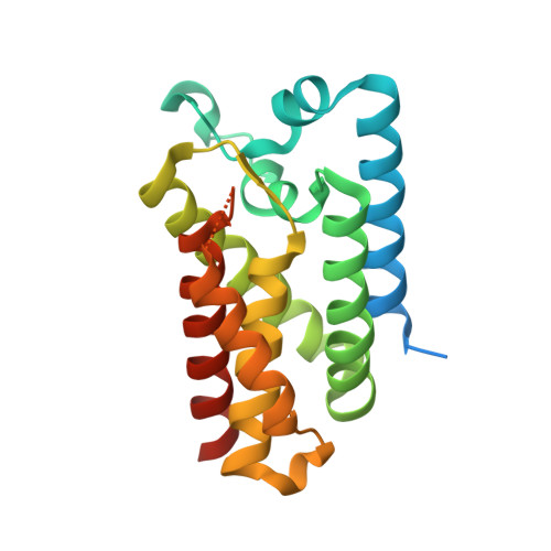
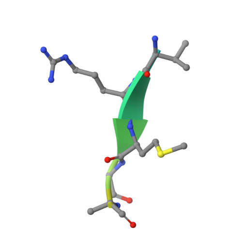
Ten catalytic snapshots of rhomboid intramembrane proteolysis from gate opening to peptide release.

Cho, S.Baker, R.P.Ji, M.Urban, S.

(2019) Nat Struct Mol Biol 26: 910-918

  • DOI: https://doi.org/10.1038/s41594-019-0296-9
  • Primary Citation of Related Structures:  
    6PJ4, 6PJ5, 6PJ7, 6PJ8, 6PJ9, 6PJA, 6PJP, 6PJQ, 6PJR, 6PJU

  • PubMed Abstract: 

    Protein cleavage inside the cell membrane triggers various pathophysiological signaling pathways, but the mechanism of catalysis is poorly understood. We solved ten structures of the Escherichia coli rhomboid protease in a bicelle membrane undergoing time-resolved steps that encompass the entire proteolytic reaction on a transmembrane substrate and an aldehyde inhibitor. Extensive gate opening accompanied substrate, but not inhibitor, binding, revealing that substrates and inhibitors take different paths to the active site. Catalysis unexpectedly commenced with, and was guided through subsequent catalytic steps by, motions of an extracellular loop, with local contributions from active site residues. We even captured the elusive tetrahedral intermediate that is uncleaved but covalently attached to the catalytic serine, about which the substrate was forced to bend dramatically. This unexpectedly stable intermediate indicates rhomboid catalysis uses an unprecedented reaction coordinate that may involve mechanically stressing the peptide bond, and could be selectively targeted by inhibitors.


  • Organizational Affiliation

    Department of Molecular Biology and Genetics, Johns Hopkins University School of Medicine, Baltimore, MD, USA.


Macromolecules
Find similar proteins by:  (by identity cutoff)  |  3D Structure
Entity ID: 1
MoleculeChains Sequence LengthOrganismDetailsImage
Rhomboid protease GlpG211Escherichia coliMutation(s): 1 
Gene Names: glpGAPT88_21985SK83_00858
EC: 3.4.21.105
Membrane Entity: Yes 
UniProt
Find proteins for P09391 (Escherichia coli (strain K12))
Explore P09391 
Go to UniProtKB:  P09391
Entity Groups  
Sequence Clusters30% Identity50% Identity70% Identity90% Identity95% Identity100% Identity
UniProt GroupP09391
Sequence Annotations
Expand
  • Reference Sequence

Find similar proteins by:  Sequence   |   3D Structure  

Entity ID: 2
MoleculeChains Sequence LengthOrganismDetailsImage
Peptide aldehyde inhibitor13Drosophila melanogasterMutation(s): 0 
Membrane Entity: Yes 
UniProt
Find proteins for P42287 (Drosophila melanogaster)
Explore P42287 
Go to UniProtKB:  P42287
Entity Groups  
Sequence Clusters30% Identity50% Identity70% Identity90% Identity95% Identity100% Identity
UniProt GroupP42287
Sequence Annotations
Expand
  • Reference Sequence
Experimental Data & Validation

Experimental Data

  • Method: X-RAY DIFFRACTION
  • Resolution: 2.50 Å
  • R-Value Free: 0.288 
  • R-Value Work: 0.241 
  • R-Value Observed: 0.243 
  • Space Group: C 2 2 21
Unit Cell:
Length ( Å )Angle ( ˚ )
a = 71.739α = 90
b = 96.45β = 90
c = 62.57γ = 90
Software Package:
Software NamePurpose
REFMACrefinement
iMOSFLMdata reduction
Aimlessdata scaling
MOLREPphasing

Structure Validation

View Full Validation Report


Entry History & Funding Information

Deposition Data


Funding OrganizationLocationGrant Number
National Institutes of Health/National Human Genome Research Institute (NIH/NHGRI)United StatesR01AI066025

Revision History  (Full details and data files)

  • Version 1.0: 2019-11-27
    Type: Initial release
  • Version 1.1: 2019-12-18
    Changes: Author supporting evidence
  • Version 1.2: 2023-10-11
    Changes: Data collection, Database references, Refinement description