6PIB

Structure of the Klebsiella pneumoniae LpxH-AZ1 complex

  • Classification: BIOSYNTHETIC PROTEIN
  • Organism(s): Klebsiella pneumoniae
  • Expression System: Escherichia coli
  • Mutation(s): No 
  • Membrane Protein: Yes  OPM

  • Deposited: 2019-06-26 Released: 2020-02-12 
  • Deposition Author(s): Cho, J., Zhou, P.
  • Funding Organization(s): National Institutes of Health/National Institute of General Medical Sciences (NIH/NIGMS), National Institutes of Health/National Institute Of Allergy and Infectious Diseases (NIH/NIAID)

Experimental Data Snapshot

  • Method: X-RAY DIFFRACTION
  • Resolution: 2.26 Å
  • R-Value Free: 0.239 
  • R-Value Work: 0.196 
  • R-Value Observed: 0.198 

wwPDB Validation   3D Report Full Report

Currently 6PIB does not have a validation slider image.


This is version 1.3 of the entry. See complete history


Literature

Structural basis of the UDP-diacylglucosamine pyrophosphohydrolase LpxH inhibition by sulfonyl piperazine antibiotics.

Cho, J.Lee, M.Cochrane, C.S.Webster, C.G.Fenton, B.A.Zhao, J.Hong, J.Zhou, P.

(2020) Proc Natl Acad Sci U S A 117: 4109-4116

  • DOI: https://doi.org/10.1073/pnas.1912876117
  • Primary Citation of Related Structures:  
    6PH9, 6PIB, 6PJ3

  • PubMed Abstract: 

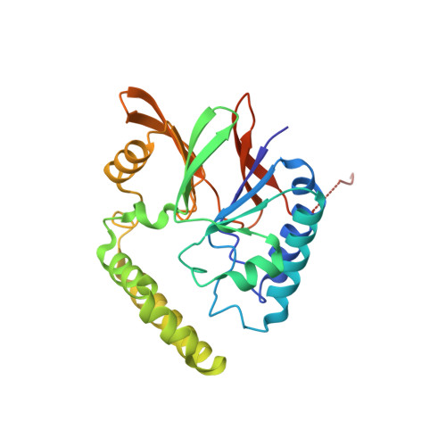
    The UDP-2,3-diacylglucosamine pyrophosphate hydrolase LpxH is an essential lipid A biosynthetic enzyme that is conserved in the majority of gram-negative bacteria. It has emerged as an attractive novel antibiotic target due to the recent discovery of an LpxH-targeting sulfonyl piperazine compound (referred to as AZ1) by AstraZeneca. However, the molecular details of AZ1 inhibition have remained unresolved, stymieing further development of this class of antibiotics. Here we report the crystal structure of Klebsiella pneumoniae LpxH in complex with AZ1. We show that AZ1 fits snugly into the L -shaped acyl chain-binding chamber of LpxH with its indoline ring situating adjacent to the active site, its sulfonyl group adopting a sharp kink, and its N -CF 3 -phenyl substituted piperazine group reaching out to the far side of the LpxH acyl chain-binding chamber. Intriguingly, despite the observation of a single AZ1 conformation in the crystal structure, our solution NMR investigation has revealed the presence of a second ligand conformation invisible in the crystalline state. Together, these distinct ligand conformations delineate a cryptic inhibitor envelope that expands the observed footprint of AZ1 in the LpxH-bound crystal structure and enables the design of AZ1 analogs with enhanced potency in enzymatic assays. These designed compounds display striking improvement in antibiotic activity over AZ1 against wild-type K. pneumoniae , and coadministration with outer membrane permeability enhancers profoundly sensitizes Escherichia coli to designed LpxH inhibitors. Remarkably, none of the sulfonyl piperazine compounds occupies the active site of LpxH, foretelling a straightforward path for rapid optimization of this class of antibiotics.


  • Organizational Affiliation

    Department of Biochemistry, Duke University Medical Center, Durham, NC 27710.


Macromolecules
Find similar proteins by:  (by identity cutoff)  |  3D Structure
Entity ID: 1
MoleculeChains Sequence LengthOrganismDetailsImage
UDP-2,3-diacylglucosamine hydrolase260Klebsiella pneumoniaeMutation(s): 0 
Gene Names: lpxH_1lpxHC3483_19950NCTC9128_00880SAMEA104305404_03891
EC: 3.6.1.54
Membrane Entity: Yes 
UniProt
Find proteins for A6T5R0 (Klebsiella pneumoniae subsp. pneumoniae (strain ATCC 700721 / MGH 78578))
Explore A6T5R0 
Go to UniProtKB:  A6T5R0
Entity Groups  
Sequence Clusters30% Identity50% Identity70% Identity90% Identity95% Identity100% Identity
UniProt GroupA6T5R0
Sequence Annotations
Expand
  • Reference Sequence
Small Molecules
Binding Affinity Annotations 
IDSourceBinding Affinity
OKV Binding MOAD:  6PIB IC50: 360 (nM) from 1 assay(s)
Experimental Data & Validation

Experimental Data

  • Method: X-RAY DIFFRACTION
  • Resolution: 2.26 Å
  • R-Value Free: 0.239 
  • R-Value Work: 0.196 
  • R-Value Observed: 0.198 
  • Space Group: P 32 2 1
Unit Cell:
Length ( Å )Angle ( ˚ )
a = 106.75α = 90
b = 106.75β = 90
c = 52.97γ = 120
Software Package:
Software NamePurpose
PHENIXrefinement
XDSdata reduction
XSCALEdata scaling
PHASERphasing

Structure Validation

View Full Validation Report

Currently 6PIB does not have a validation slider image.


Entry History & Funding Information

Deposition Data


Funding OrganizationLocationGrant Number
National Institutes of Health/National Institute of General Medical Sciences (NIH/NIGMS)United StatesGM115355
National Institutes of Health/National Institute Of Allergy and Infectious Diseases (NIH/NIAID)United StatesAI139216

Revision History  (Full details and data files)

  • Version 1.0: 2020-02-12
    Type: Initial release
  • Version 1.1: 2020-02-26
    Changes: Database references
  • Version 1.2: 2020-03-11
    Changes: Database references
  • Version 1.3: 2023-10-11
    Changes: Data collection, Database references, Refinement description