6PHA

Protein Tyrosine Phosphatase 1B (1-301), F182A mutant, vanadate bound state


Experimental Data Snapshot

  • Method: X-RAY DIFFRACTION
  • Resolution: 2.30 Å
  • R-Value Free: 0.277 
  • R-Value Work: 0.224 
  • R-Value Observed: 0.227 

wwPDB Validation   3D Report Full Report


This is version 1.4 of the entry. See complete history


Literature

Uncovering the Molecular Interactions in the Catalytic Loop That Modulate the Conformational Dynamics in Protein Tyrosine Phosphatase 1B.

Cui, D.S.Lipchock, J.M.Brookner, D.Loria, J.P.

(2019) J Am Chem Soc 141: 12634-12647

  • DOI: https://doi.org/10.1021/jacs.9b04470
  • Primary Citation of Related Structures:  
    6OL4, 6OLQ, 6OLV, 6OMY, 6PFW, 6PG0, 6PGT, 6PHA, 6PHS, 6PM8

  • PubMed Abstract: 

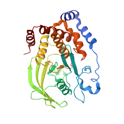
    Active-site loops are integral to the function of numerous enzymes. They enable substrate and product binding and release, sequester reaction intermediates, and recruit catalytic groups. Here, we examine the catalytic loop in the enzyme protein tyrosine phosphatase 1B (PTP1B). PTP1B has a mobile so-called WPD loop (named for its three N-terminal residues) that initiates the dephosphorylation of phosphor-tyrosine substrates upon loop closure. We have combined X-ray crystallography, solution NMR, and pre-steady-state kinetics experiments on wild-type and five WPD loop mutants to identify the relationships between the loop structure, dynamics, and function. The motions of the WPD loop are modulated by the formation of weak molecular interactions, where perturbations of these interactions modulate the conformational equilibrium landscape. The point mutants in the WPD loop alter the loop equilibrium position from a predominantly open state (P185A) to 50:50 (F182A), 35:65 (P188A), and predominantly closed states (T177A and P188A). Surprisingly, there is no correlation between the observed catalytic rates in the loop mutants and changes to the WPD loop equilibrium position. Rather, we observe a strong correlation between the rate of dephosphorylation of the phosphocysteine enzyme intermediate and uniform millisecond motions, not only within the loop but also in the adjacent α-helical domain of PTP1B. Thus, the control of loop motion and thereby catalytic activity is dispersed and resides within not only the loop sequence but also the surrounding protein architecture. This has broad implications for the general mechanistic understanding of enzyme reactions and the role that flexible loops play in the catalytic cycle.


  • Organizational Affiliation

    Department of Chemistry , Yale University , New Haven , Connecticut 06511 , United States.


Macromolecules
Find similar proteins by:  (by identity cutoff)  |  3D Structure
Entity ID: 1
MoleculeChains Sequence LengthOrganismDetailsImage
Tyrosine-protein phosphatase non-receptor type 1297Homo sapiensMutation(s): 1 
Gene Names: PTPN1PTP1B
EC: 3.1.3.48
UniProt & NIH Common Fund Data Resources
Find proteins for P18031 (Homo sapiens)
Explore P18031 
Go to UniProtKB:  P18031
PHAROS:  P18031
GTEx:  ENSG00000196396 
Entity Groups  
Sequence Clusters30% Identity50% Identity70% Identity90% Identity95% Identity100% Identity
UniProt GroupP18031
Sequence Annotations
Expand
  • Reference Sequence
Small Molecules
Ligands 3 Unique
IDChains Name / Formula / InChI Key2D Diagram3D Interactions
VO4 (Subject of Investigation/LOI)
Query on VO4

Download Ideal Coordinates CCD File 
B [auth A]VANADATE ION
O4 V
LSGOVYNHVSXFFJ-UHFFFAOYSA-N
GOL
Query on GOL

Download Ideal Coordinates CCD File 
C [auth A],
D [auth A],
E [auth A],
F [auth A],
G [auth A]
GLYCEROL
C3 H8 O3
PEDCQBHIVMGVHV-UHFFFAOYSA-N
ACT
Query on ACT

Download Ideal Coordinates CCD File 
H [auth A],
I [auth A],
J [auth A]
ACETATE ION
C2 H3 O2
QTBSBXVTEAMEQO-UHFFFAOYSA-M
Binding Affinity Annotations 
IDSourceBinding Affinity
VO4 BindingDB:  6PHA IC50: min: 25.39, max: 1.52e+4 (nM) from 12 assay(s)
Experimental Data & Validation

Experimental Data

  • Method: X-RAY DIFFRACTION
  • Resolution: 2.30 Å
  • R-Value Free: 0.277 
  • R-Value Work: 0.224 
  • R-Value Observed: 0.227 
  • Space Group: P 31 2 1
Unit Cell:
Length ( Å )Angle ( ˚ )
a = 87.56α = 90
b = 87.56β = 90
c = 102.376γ = 120
Software Package:
Software NamePurpose
PHENIXrefinement
HKL-2000data reduction
HKL-2000data scaling
PHASERphasing

Structure Validation

View Full Validation Report


Entry History & Funding Information

Deposition Data


Funding OrganizationLocationGrant Number
National Institutes of Health/National Institute of General Medical Sciences (NIH/NIGMS)United StatesRO1GM112781
National Science Foundation (NSF, United States)United StatesMCB1615415

Revision History  (Full details and data files)

  • Version 1.0: 2019-08-07
    Type: Initial release
  • Version 1.1: 2019-08-28
    Changes: Data collection, Database references
  • Version 1.2: 2019-11-27
    Changes: Author supporting evidence
  • Version 1.3: 2020-03-25
    Changes: Database references
  • Version 1.4: 2023-10-11
    Changes: Data collection, Database references, Refinement description