6PGL

Structure of Kluyveromyces marxianus Usb1 with uridine monophosphate


Experimental Data Snapshot

  • Method: X-RAY DIFFRACTION
  • Resolution: 1.84 Å
  • R-Value Free: 0.219 
  • R-Value Work: 0.188 
  • R-Value Observed: 0.189 

wwPDB Validation   3D Report Full Report


Ligand Structure Quality Assessment 


This is version 1.2 of the entry. See complete history


Literature

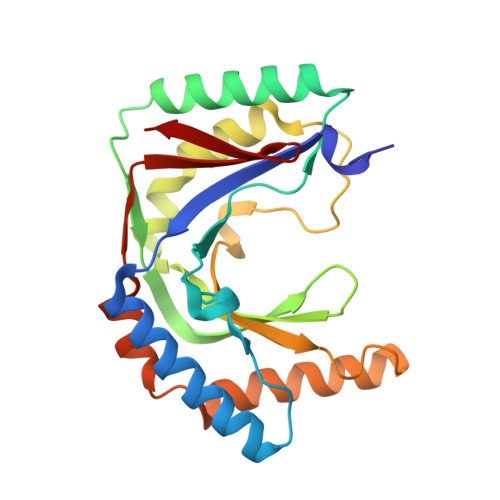
Structural basis for the evolution of cyclic phosphodiesterase activity in the U6 snRNA exoribonuclease Usb1.

Nomura, Y.Montemayor, E.J.Virta, J.M.Hayes, S.M.Butcher, S.E.

(2020) Nucleic Acids Res 48: 1423-1434

  • DOI: https://doi.org/10.1093/nar/gkz1177
  • Primary Citation of Related Structures:  
    6PFQ, 6PGL

  • PubMed Abstract: 

    U6 snRNA undergoes post-transcriptional 3' end modification prior to incorporation into the active site of spliceosomes. The responsible exoribonuclease is Usb1, which removes nucleotides from the 3' end of U6 and, in humans, leaves a 2',3' cyclic phosphate that is recognized by the Lsm2-8 complex. Saccharomycescerevisiae Usb1 has additional 2',3' cyclic phosphodiesterase (CPDase) activity, which converts the cyclic phosphate into a 3' phosphate group. Here we investigate the molecular basis for the evolution of Usb1 CPDase activity. We examine the structure and function of Usb1 from Kluyveromyces marxianus, which shares 25 and 19% sequence identity to the S. cerevisiae and Homo sapiens orthologs of Usb1, respectively. We show that K. marxianus Usb1 enzyme has CPDase activity and determined its structure, free and bound to the substrate analog uridine 5'-monophosphate. We find that the origin of CPDase activity is related to a loop structure that is conserved in yeast and forms a distinct penultimate (n - 1) nucleotide binding site. These data provide structural and mechanistic insight into the evolutionary divergence of Usb1 catalysis.


  • Organizational Affiliation

    Department of Biochemistry, University of Wisconsin, Madison, WI 53706, USA.


Macromolecules
Find similar proteins by:  (by identity cutoff)  |  3D Structure
Entity ID: 1
MoleculeChains Sequence LengthOrganismDetailsImage
Uncharacterized protein YLR132C
A, B, C
218Kluyveromyces marxianus DMKU3-1042Mutation(s): 0 
Gene Names: KLMA_50491
UniProt
Find proteins for W0TCJ5 (Kluyveromyces marxianus (strain DMKU3-1042 / BCC 29191 / NBRC 104275))
Explore W0TCJ5 
Go to UniProtKB:  W0TCJ5
Entity Groups  
Sequence Clusters30% Identity50% Identity70% Identity90% Identity95% Identity100% Identity
UniProt GroupW0TCJ5
Sequence Annotations
Expand
  • Reference Sequence
Small Molecules
Ligands 6 Unique
IDChains Name / Formula / InChI Key2D Diagram3D Interactions
U5P (Subject of Investigation/LOI)
Query on U5P

Download Ideal Coordinates CCD File 
E [auth A],
I [auth B],
P [auth C]
URIDINE-5'-MONOPHOSPHATE
C9 H13 N2 O9 P
DJJCXFVJDGTHFX-XVFCMESISA-N
PG6
Query on PG6

Download Ideal Coordinates CCD File 
N [auth B],
V [auth C]
1-(2-METHOXY-ETHOXY)-2-{2-[2-(2-METHOXY-ETHOXY]-ETHOXY}-ETHANE
C12 H26 O6
DMDPGPKXQDIQQG-UHFFFAOYSA-N
PG4
Query on PG4

Download Ideal Coordinates CCD File 
U [auth C]TETRAETHYLENE GLYCOL
C8 H18 O5
UWHCKJMYHZGTIT-UHFFFAOYSA-N
PGE
Query on PGE

Download Ideal Coordinates CCD File 
F [auth A],
G [auth A],
H [auth A],
J [auth B],
Q [auth C]
TRIETHYLENE GLYCOL
C6 H14 O4
ZIBGPFATKBEMQZ-UHFFFAOYSA-N
GOL
Query on GOL

Download Ideal Coordinates CCD File 
D [auth A],
O [auth C]
GLYCEROL
C3 H8 O3
PEDCQBHIVMGVHV-UHFFFAOYSA-N
EDO
Query on EDO

Download Ideal Coordinates CCD File 
K [auth B]
L [auth B]
M [auth B]
R [auth C]
S [auth C]
K [auth B],
L [auth B],
M [auth B],
R [auth C],
S [auth C],
T [auth C]
1,2-ETHANEDIOL
C2 H6 O2
LYCAIKOWRPUZTN-UHFFFAOYSA-N
Experimental Data & Validation

Experimental Data

  • Method: X-RAY DIFFRACTION
  • Resolution: 1.84 Å
  • R-Value Free: 0.219 
  • R-Value Work: 0.188 
  • R-Value Observed: 0.189 
  • Space Group: P 21 21 21
Unit Cell:
Length ( Å )Angle ( ˚ )
a = 69.957α = 90
b = 110.344β = 90
c = 114.697γ = 90
Software Package:
Software NamePurpose
PHENIXrefinement
STARANISOdata scaling
PHENIXphasing

Structure Validation

View Full Validation Report


Ligand Structure Quality Assessment 


Entry History & Funding Information

Deposition Data


Funding OrganizationLocationGrant Number
National Institutes of Health/National Human Genome Research Institute (NIH/NHGRI)United StatesR01 GM065166
National Institutes of Health/National Human Genome Research Institute (NIH/NHGRI)United StatesR35 GM118131

Revision History  (Full details and data files)

  • Version 1.0: 2019-12-11
    Type: Initial release
  • Version 1.1: 2020-05-20
    Changes: Database references
  • Version 1.2: 2024-03-13
    Changes: Data collection, Database references