6PAY | pdb_00006pay

Structure of HsICDH1:Mg(II):ICT:NADPH(50%) complex reveals structural basis for observation of half-sites reactivity


Experimental Data Snapshot

  • Method: X-RAY DIFFRACTION
  • Resolution: 2.20 Å
  • R-Value Free: 
    0.242 (Depositor), 0.244 (DCC) 
  • R-Value Work: 
    0.224 (Depositor), 0.227 (DCC) 
  • R-Value Observed: 
    0.225 (Depositor) 

Starting Model: experimental
View more details

wwPDB Validation 3D Report Full Report

Validation slider image for 6PAY

Ligand Structure Quality Assessment 


This is version 1.2 of the entry. See complete history

Literature

Transient-State Analysis of Human Isocitrate Dehydrogenase I: Accounting for the Interconversion of Active and Non-Active Conformational States.

Roman, J.V.Melkonian, T.R.Silvaggi, N.R.Moran, G.R.

(2019) Biochemistry 58: 5366-5380

  • DOI: https://doi.org/10.1021/acs.biochem.9b00518
  • Primary Citation Related Structures: 
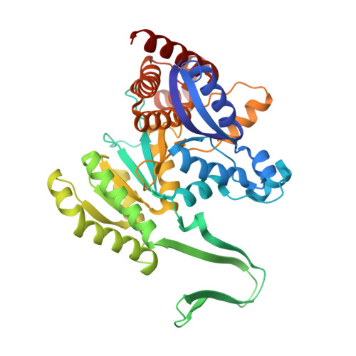
    6PAY

  • PubMed Abstract: 

    Human isocitrate dehydrogenase 1 (HsICDH1) is a cytoplasmic homodimeric Mg(II)-dependent enzyme that converts d-isocitrate (D-ICT) and NADP + to α-ketoglutarate (AKG), CO 2 , and NADPH. The active sites are formed at the subunit interface and incorporate residues from both protomers. The turnover number titrates hyperbolically from 17.5 s -1 to a minimum of 7 s -1 with an increasing enzyme concentration. As isolated, the enzyme adopts an inactive open conformation and binds NADPH tightly. The open conformation displaces three of the eight residues that bind D-ICT and Mg(II). Enzyme activation occurs with the addition of Mg(II) or D-ICT with a rate constant of 0.12 s -1 . The addition of both Mg(II) and D-ICT activates the enzyme with a rate constant of 0.6 s -1 and displaces half of the bound NADPH. This indicates that HsICDH1 may have a half-site mechanism in which the active sites alternate in catalysis. The X-ray crystal structure of the half-site activated complex reveals asymmetry in the homodimer with a single NADPH bound. The structure also indicates a pseudotetramer interface that impedes the egress of NADPH consistent with the suppression of the turnover number at high enzyme concentrations. When the half-site activated form of the enzyme is reacted with NADP + , NADPH forms with a rate constant of 204 s -1 followed by a shift in the NADPH absorption spectrum with a rate constant of 28 s -1 . These data indicate the accumulation of two intermediate states. Once D-ICT is exhausted, HsICDH1 relaxes to the inactive open state with a rate constant of ∼3 s -1 .


  • Organizational Affiliation
    • Department of Chemistry and Biochemistry , Loyola University Chicago , Flanner Hall, 1068 West Sheridan Road , Chicago , Illinois 60660 , United States.

Macromolecule Content 

  • Total Structure Weight: 188.81 kDa 
  • Atom Count: 13,235 
  • Modeled Residue Count: 1,635 
  • Deposited Residue Count: 1,656 
  • Unique protein chains: 1

Macromolecules

Find similar proteins by:|  3D Structure
Entity ID: 1
MoleculeChains  Sequence LengthOrganismDetailsImage
Isocitrate dehydrogenase [NADP] cytoplasmic
A, B, C, D
414Homo sapiensMutation(s): 0 
Gene Names: IDH1PICD
EC: 1.1.1.42
UniProt & NIH Common Fund Data Resources
Find proteins for O75874 (Homo sapiens)
Explore O75874 
Go to UniProtKB:  O75874
PHAROS:  O75874
GTEx:  ENSG00000138413 
Entity Groups
Sequence Clusters30% Identity50% Identity70% Identity90% Identity95% Identity100% Identity
UniProt GroupO75874
Sequence Annotations
Expand
Reference Sequence

Small Molecules

Ligands 4 Unique
IDChains Name / Formula / InChI Key2D Diagram3D Interactions
NDP

Query on NDP


Download:Ideal Coordinates CCD File
G [auth A]NADPH DIHYDRO-NICOTINAMIDE-ADENINE-DINUCLEOTIDE PHOSPHATE
C21 H30 N7 O17 P3
ACFIXJIJDZMPPO-NNYOXOHSSA-N
ICT

Query on ICT


Download:Ideal Coordinates CCD File
E [auth A],
L [auth B],
P [auth C],
R [auth D]
ISOCITRIC ACID
C6 H8 O7
ODBLHEXUDAPZAU-ZAFYKAAXSA-N
FMT

Query on FMT


Download:Ideal Coordinates CCD File
H [auth A]
I [auth A]
J [auth A]
K [auth A]
N [auth B]
H [auth A],
I [auth A],
J [auth A],
K [auth A],
N [auth B],
O [auth B],
T [auth D]
FORMIC ACID
C H2 O2
BDAGIHXWWSANSR-UHFFFAOYSA-N
MG

Query on MG


Download:Ideal Coordinates CCD File
F [auth A],
M [auth B],
Q [auth C],
S [auth D]
MAGNESIUM ION
Mg
JLVVSXFLKOJNIY-UHFFFAOYSA-N

Experimental Data & Validation

Experimental Data

  • Method: X-RAY DIFFRACTION
  • Resolution: 2.20 Å
  • R-Value Free:  0.242 (Depositor), 0.244 (DCC) 
  • R-Value Work:  0.224 (Depositor), 0.227 (DCC) 
  • R-Value Observed: 0.225 (Depositor) 
Space Group: P 1 21 1
Unit Cell:
Length ( Å )Angle ( ˚ )
a = 94.6α = 90
b = 74.6β = 95.2
c = 142.9γ = 90
Software Package:
Software NamePurpose
PHENIXrefinement
HKL-2000data reduction
HKL-2000data scaling
PHASERphasing

Structure Validation

View Full Validation Report


Ligand Structure Quality Assessment 


Entry History 

Deposition Data

Revision History  (Full details and data files)

  • Version 1.0: 2019-09-18
    Type: Initial release
  • Version 1.1: 2020-01-08
    Changes: Database references
  • Version 1.2: 2023-10-11
    Changes: Data collection, Database references, Derived calculations, Refinement description