6PAK | pdb_00006pak

Insight into subtilisin E-S7 cleavage pattern based on crystal structure and hydrolysates peptide analysis


Experimental Data Snapshot

  • Method: X-RAY DIFFRACTION
  • Resolution: 1.98 Å
  • R-Value Free: 
    0.286 (Depositor), 0.290 (DCC) 
  • R-Value Work: 
    0.239 (Depositor), 0.240 (DCC) 
  • R-Value Observed: 
    0.242 (Depositor) 

Starting Model: experimental
View more details

wwPDB Validation 3D Report Full Report

Validation slider image for 6PAK

This is version 1.5 of the entry. See complete history

Literature

Enhancing subtilisin thermostability through a modified normalized B-factor analysis and loop-grafting strategy.

Tang, H.Shi, K.Shi, C.Aihara, H.Zhang, J.Du, G.

(2019) J Biological Chem 294: 18398-18407

  • DOI: https://doi.org/10.1074/jbc.RA119.010658
  • Primary Citation Related Structures: 
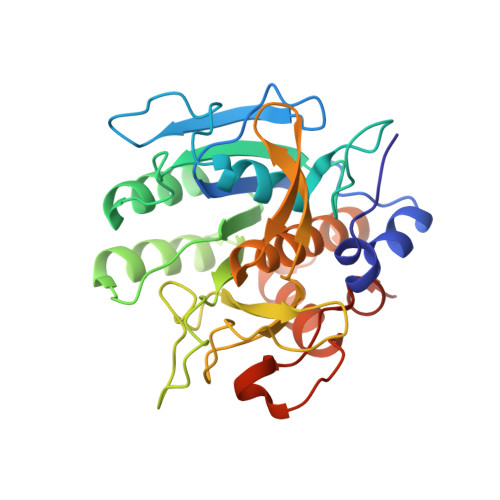
    6PAK

  • PubMed Abstract: 

    Rational design-guided improvement of protein thermostability typically requires identification of residues or regions contributing to instability and introduction of mutations into these residues or regions. One popular method, B-FIT, utilizes B-factors to identify unstable residues or regions and combines them with other strategies, such as directed evolution. Here, we performed structure-based engineering to improve the thermostability of the subtilisin E-S7 (SES7) peptidase. The B-value of each residue was redefined in a normalized B-factor calculation, which was implemented with a refined bioinformatics analysis strategy to identify the critical area (loop 158-162) related to flexibility and to screen for suitable thermostable motif sequences in the Protein Data Bank that can act as transplant loops. In total, we analyzed 445 structures and identified 29 thermostable motifs as candidates. Using these motifs as a starting point, we performed iterative homologous modeling to obtain a desirable chimera loop and introduced five different mutations into this loop to construct thermostable SES7 proteins. Differential scanning fluorimetry revealed increases of 7.3 °C in the melting temperature of an SES7 variant designated M5 compared with the WT. The X-ray crystallographic structure of this variant was resolved at 1.96 Å resolution. The crystal structure disclosed that M5 forms more hydrogen bonds than the WT protein, consistent with design and molecular dynamics simulation results. In summary, the modified B-FIT strategy reported here has yielded a subtilisin variant with improved thermostability and promising industrial applications, supporting the notion that this modified method is a powerful tool for protein engineering.


  • Organizational Affiliation
    • Key Laboratory of Industrial Biotechnology, Ministry of Education, Jiangnan University, 1800 Lihu Road, Wuxi 214122, Jiangsu, China; School of Biotechnology, Ministry of Education, Jiangnan University, 1800 Lihu Road, Wuxi 214122, Jiangsu, China.

Macromolecule Content 

  • Total Structure Weight: 58.6 kDa 
  • Atom Count: 4,420 
  • Modeled Residue Count: 554 
  • Deposited Residue Count: 570 
  • Unique protein chains: 1

Macromolecules

Find similar proteins by:|  3D Structure
Entity ID: 1
MoleculeChains  Sequence LengthOrganismDetailsImage
Subtilisin E
A, B
285Bacillus subtilis subsp. subtilis str. 168Mutation(s): 7 
Gene Names: aprEapraprAsprEBSU10300
EC: 3.4.21.62
UniProt
Find proteins for P04189 (Bacillus subtilis (strain 168))
Explore P04189 
Go to UniProtKB:  P04189
Entity Groups
Sequence Clusters30% Identity50% Identity70% Identity90% Identity95% Identity100% Identity
UniProt GroupP04189
Sequence Annotations
Expand
Reference Sequence

Experimental Data & Validation

Experimental Data

  • Method: X-RAY DIFFRACTION
  • Resolution: 1.98 Å
  • R-Value Free:  0.286 (Depositor), 0.290 (DCC) 
  • R-Value Work:  0.239 (Depositor), 0.240 (DCC) 
  • R-Value Observed: 0.242 (Depositor) 
Space Group: P 21 21 21
Unit Cell:
Length ( Å )Angle ( ˚ )
a = 74.247α = 90
b = 80.475β = 90
c = 86.92γ = 90
Software Package:
Software NamePurpose
PHENIXrefinement
XDSdata reduction
Aimlessdata scaling
PHENIXphasing

Structure Validation

View Full Validation Report


Entry History 

& Funding Information

Deposition Data


Funding OrganizationLocationGrant Number
National Institutes of Health/National Institute of General Medical Sciences (NIH/NIGMS)United StatesGM118047
National Natural Science Foundation of China (NSFC)China31470160

Revision History  (Full details and data files)

  • Version 1.0: 2019-10-23
    Type: Initial release
  • Version 1.1: 2019-10-30
    Changes: Data collection, Database references
  • Version 1.2: 2019-12-11
    Changes: Database references
  • Version 1.3: 2019-12-18
    Changes: Database references
  • Version 1.4: 2020-01-01
    Changes: Author supporting evidence
  • Version 1.5: 2023-10-11
    Changes: Data collection, Database references, Refinement description