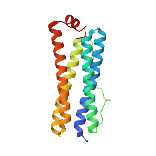
6P8K

Escherichia coli Bacterioferritin Substituted with Zinc Protoporphyrin IX


Experimental Data Snapshot

  • Method: X-RAY DIFFRACTION
  • Resolution: 1.70 Å
  • R-Value Free: 0.217 
  • R-Value Work: 0.192 
  • R-Value Observed: 0.192 

wwPDB Validation   3D Report Full Report


Ligand Structure Quality Assessment 


This is version 1.1 of the entry. See complete history


Literature

Structure of a Zinc Porphyrin-Substituted Bacterioferritin and Photophysical Properties of Iron Reduction.

Benavides, B.S.Valandro, S.Cioloboc, D.Taylor, A.B.Schanze, K.S.Kurtz Jr., D.M.

(2020) Biochemistry 59: 1618-1629

  • DOI: https://doi.org/10.1021/acs.biochem.9b01103
  • Primary Citation of Related Structures:  
    6P8K, 6P8L

  • PubMed Abstract: 

    The iron storage protein bacterioferritin (Bfr) binds up to 12 hemes b at specific sites in its protein shell. The heme b can be substituted with the photosensitizer Zn(II)-protoporphyrin IX (ZnPP), and photosensitized reductive iron release from the ferric oxyhydroxide {[FeO(OH)] n } core inside the ZnPP-Bfr protein shell was demonstrated [Cioloboc, D., et al. (2018) Biomacromolecules 19 , 178-187]. This report describes the X-ray crystal structure of ZnPP-Bfr and the effects of loaded iron on the photophysical properties of the ZnPP. The crystal structure of ZnPP-Bfr shows a unique six-coordinate zinc in the ZnPP with two axial methionine sulfur ligands. Steady state and transient ultraviolet-visible absorption and luminescence spectroscopies show that irradiation with light overlapping the Soret absorption causes oxidation of ZnPP to the cation radical ZnPP •+ only when the ZnPP-Bfr is loaded with [FeO(OH)] n . Femtosecond transient absorption spectroscopy shows that this photooxidation occurs from the singlet excited state ( 1 ZnPP*) on the picosecond time scale and is consistent with two oxidizing populations of Fe 3+ , which do not appear to involve the ferroxidase center iron. We propose that [FeO(OH)] n clusters at or near the inner surface of the protein shell are responsible for ZnPP photooxidation. Hopping of the photoinjected electrons through the [FeO(OH)] n would effectively cause migration of Fe 2+ through the inner cavity to pores where it exits the protein. Reductive iron mobilization is presumed to be a physiological function of Bfrs. The phototriggered Fe 3+ reduction could be used to identify the sites of iron mobilization within the Bfr protein shell.


  • Organizational Affiliation

    Department of Chemistry, University of Texas at San Antonio, San Antonio, Texas 78249, United States.


Macromolecules
Find similar proteins by:  (by identity cutoff)  |  3D Structure
Entity ID: 1
MoleculeChains Sequence LengthOrganismDetailsImage
Bacterioferritin
A, B, C, D, E
A, B, C, D, E, F, G, H, I, J, K, L
158Escherichia coli K-12Mutation(s): 0 
Gene Names: bfrb3336JW3298
EC: 1.16.3.1
UniProt
Find proteins for P0ABD3 (Escherichia coli (strain K12))
Explore P0ABD3 
Go to UniProtKB:  P0ABD3
Entity Groups  
Sequence Clusters30% Identity50% Identity70% Identity90% Identity95% Identity100% Identity
UniProt GroupP0ABD3
Sequence Annotations
Expand
  • Reference Sequence
Small Molecules
Ligands 4 Unique
IDChains Name / Formula / InChI Key2D Diagram3D Interactions
ZNH (Subject of Investigation/LOI)
Query on ZNH

Download Ideal Coordinates CCD File 
DB [auth K]
GA [auth E]
N [auth A]
OA [auth G]
Y [auth C]
DB [auth K],
GA [auth E],
N [auth A],
OA [auth G],
Y [auth C],
YA [auth J]
PROTOPORPHYRIN IX CONTAINING ZN
C34 H32 N4 O4 Zn
FUTVBRXUIKZACV-RGGAHWMASA-L
MLI
Query on MLI

Download Ideal Coordinates CCD File 
AA [auth C]
AB [auth J]
BB [auth J]
DA [auth D]
EA [auth D]
AA [auth C],
AB [auth J],
BB [auth J],
DA [auth D],
EA [auth D],
EB [auth K],
FB [auth K],
HA [auth E],
HB [auth L],
IA [auth E],
IB [auth L],
JB [auth L],
LA [auth F],
MA [auth F],
P [auth A],
PA [auth G],
Q [auth A],
QA [auth G],
R [auth A],
SA [auth H],
TA [auth H],
U [auth B],
V [auth B],
VA [auth I],
W [auth B],
WA [auth I],
XA [auth I],
Z [auth C]
MALONATE ION
C3 H2 O4
OFOBLEOULBTSOW-UHFFFAOYSA-L
ZN
Query on ZN

Download Ideal Coordinates CCD File 
BA [auth D]
CB [auth K]
FA [auth E]
GB [auth L]
JA [auth F]
BA [auth D],
CB [auth K],
FA [auth E],
GB [auth L],
JA [auth F],
M [auth A],
NA [auth G],
RA [auth H],
S [auth B],
UA [auth I],
X [auth C],
ZA [auth J]
ZINC ION
Zn
PTFCDOFLOPIGGS-UHFFFAOYSA-N
NA
Query on NA

Download Ideal Coordinates CCD File 
CA [auth D],
KA [auth F],
O [auth A],
T [auth B]
SODIUM ION
Na
FKNQFGJONOIPTF-UHFFFAOYSA-N
Experimental Data & Validation

Experimental Data

  • Method: X-RAY DIFFRACTION
  • Resolution: 1.70 Å
  • R-Value Free: 0.217 
  • R-Value Work: 0.192 
  • R-Value Observed: 0.192 
  • Space Group: P 42 21 2
Unit Cell:
Length ( Å )Angle ( ˚ )
a = 207.852α = 90
b = 207.852β = 90
c = 142.657γ = 90
Software Package:
Software NamePurpose
PHENIXrefinement
XDSdata reduction
XDSdata scaling
PHASERphasing

Structure Validation

View Full Validation Report


Ligand Structure Quality Assessment 


Entry History & Funding Information

Deposition Data


Funding OrganizationLocationGrant Number
National Institutes of Health/National Center for Advancing Translational Sciences (NIH/NCATS)United States8UL1TR001120

Revision History  (Full details and data files)

  • Version 1.0: 2020-05-13
    Type: Initial release
  • Version 1.1: 2023-10-11
    Changes: Data collection, Database references, Derived calculations, Refinement description