6P7G | pdb_00006p7g

The co-crystal structure of BRAF(V600E) with PHI1


Experimental Data Snapshot

  • Method: X-RAY DIFFRACTION
  • Resolution: 2.65 Å
  • R-Value Free: 
    0.279 (Depositor), 0.283 (DCC) 
  • R-Value Work: 
    0.248 (Depositor), 0.251 (DCC) 
  • R-Value Observed: 
    0.249 (Depositor) 

wwPDB Validation 3D Report Full Report

Validation slider image for 6P7G

Ligand Structure Quality Assessment 


This is version 1.1 of the entry. See complete history

Literature

Inhibitors of BRAF dimers using an allosteric site.

Cotto-Rios, X.M.Agianian, B.Gitego, N.Zacharioudakis, E.Giricz, O.Wu, Y.Zou, Y.Verma, A.Poulikakos, P.I.Gavathiotis, E.

(2020) Nat Commun 11: 4370-4370

  • DOI: https://doi.org/10.1038/s41467-020-18123-2
  • Primary Citation Related Structures: 
    6P3D, 6P7G

  • PubMed Abstract: 

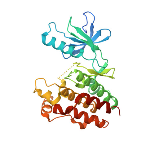
    BRAF kinase, a critical effector of the ERK signaling pathway, is hyperactivated in many cancers. Oncogenic BRAF V600E signals as an active monomer in the absence of active RAS, however, in many tumors BRAF dimers mediate ERK signaling. FDA-approved RAF inhibitors poorly inhibit BRAF dimers, which leads to tumor resistance. We found that Ponatinib, an FDA-approved drug, is an effective inhibitor of BRAF monomers and dimers. Ponatinib binds the BRAF dimer and stabilizes a distinct αC-helix conformation through interaction with a previously unrevealed allosteric site. Using these structural insights, we developed PHI1, a BRAF inhibitor that fully uncovers the allosteric site. PHI1 exhibits discrete cellular selectivity for BRAF dimers, with enhanced inhibition of the second protomer when the first protomer is occupied, comprising a novel class of dimer selective inhibitors. This work shows that Ponatinib and BRAF dimer selective inhibitors will be useful in treating BRAF-dependent tumors.


  • Organizational Affiliation
    • Department of Biochemistry, Albert Einstein College of Medicine, Bronx, NY, USA.

Macromolecule Content 

  • Total Structure Weight: 137.47 kDa 
  • Atom Count: 8,543 
  • Modeled Residue Count: 1,039 
  • Deposited Residue Count: 1,188 
  • Unique protein chains: 1

Macromolecules

Find similar proteins by:|  3D Structure
Entity ID: 1
MoleculeChains  Sequence LengthOrganismDetailsImage
Serine/threonine-protein kinase B-raf
A, B, C, D
297Homo sapiensMutation(s): 17 
Gene Names: BRAFBRAF1RAFB1
EC: 2.7.11.1
UniProt & NIH Common Fund Data Resources
Find proteins for P15056 (Homo sapiens)
Explore P15056 
Go to UniProtKB:  P15056
PHAROS:  P15056
GTEx:  ENSG00000157764 
Entity Groups
Sequence Clusters30% Identity50% Identity70% Identity90% Identity95% Identity100% Identity
UniProt GroupP15056
Sequence Annotations
Expand
Reference Sequence

Experimental Data & Validation

Experimental Data

  • Method: X-RAY DIFFRACTION
  • Resolution: 2.65 Å
  • R-Value Free:  0.279 (Depositor), 0.283 (DCC) 
  • R-Value Work:  0.248 (Depositor), 0.251 (DCC) 
  • R-Value Observed: 0.249 (Depositor) 
Space Group: P 21 21 21
Unit Cell:
Length ( Å )Angle ( ˚ )
a = 95.78α = 90
b = 108.79β = 90
c = 126.38γ = 90
Software Package:
Software NamePurpose
REFMACrefinement
SCALAdata scaling
PHASERphasing
PDB_EXTRACTdata extraction
MOSFLMdata reduction

Structure Validation

View Full Validation Report


Ligand Structure Quality Assessment 


Entry History 

& Funding Information

Deposition Data


Funding OrganizationLocationGrant Number
National Institutes of Health/National Cancer Institute (NIH/NCI)United StatesR01CA178394
National Institutes of Health/National Cancer Institute (NIH/NCI)United StatesR01CA204314
National Institutes of Health/National Cancer Institute (NIH/NCI)United StatesP30CA013330

Revision History  (Full details and data files)

  • Version 1.0: 2020-09-23
    Type: Initial release
  • Version 1.1: 2024-03-13
    Changes: Data collection, Database references, Refinement description, Structure summary