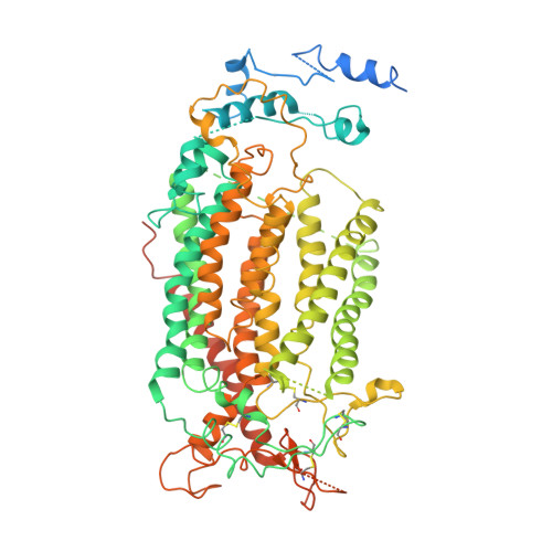
6P49

Cryo-EM structure of calcium-bound TMEM16F in nanodisc with supplement of PIP2 in Cl2


Experimental Data Snapshot

  • Method: ELECTRON MICROSCOPY
  • Resolution: 3.30 Å
  • Aggregation State: PARTICLE 
  • Reconstruction Method: SINGLE PARTICLE 

wwPDB Validation   3D Report Full Report


This is version 1.1 of the entry. See complete history


Literature

Cryo-EM Studies of TMEM16F Calcium-Activated Ion Channel Suggest Features Important for Lipid Scrambling.

Feng, S.Dang, S.Han, T.W.Ye, W.Jin, P.Cheng, T.Li, J.Jan, Y.N.Jan, L.Y.Cheng, Y.

(2019) Cell Rep 28: 567-579.e4

  • DOI: https://doi.org/10.1016/j.celrep.2019.06.023
  • Primary Citation of Related Structures:  
    6P46, 6P47, 6P48, 6P49

  • PubMed Abstract: 

    As a Ca 2+ -activated lipid scramblase and ion channel that mediates Ca 2+ influx, TMEM16F relies on both functions to facilitate extracellular vesicle generation, blood coagulation, and bone formation. How a bona fide ion channel scrambles lipids remains elusive. Our structural analyses revealed the coexistence of an intact channel pore and PIP 2 -dependent protein conformation changes leading to membrane distortion. Correlated to the extent of membrane distortion, many tightly bound lipids are slanted. Structure-based mutagenesis studies further reveal that neutralization of some lipid-binding residues or those near membrane distortion specifically alters the onset of lipid scrambling, but not Ca 2+ influx, thus identifying features outside of channel pore that are important for lipid scrambling. Together, our studies demonstrate that membrane distortion does not require open hydrophilic grooves facing the membrane interior and provide further evidence to suggest separate pathways for lipid scrambling and ion permeation.


  • Organizational Affiliation

    Department of Physiology, University of California, San Francisco, San Francisco, CA 94158, USA.


Macromolecules
Find similar proteins by:  (by identity cutoff)  |  3D Structure
Entity ID: 1
MoleculeChains Sequence LengthOrganismDetailsImage
Anoctamin-6
A, B
911Mus musculusMutation(s): 0 
Gene Names: Ano6Tmem16f
Membrane Entity: Yes 
UniProt & NIH Common Fund Data Resources
Find proteins for Q6P9J9 (Mus musculus)
Explore Q6P9J9 
Go to UniProtKB:  Q6P9J9
IMPC:  MGI:2145890
Entity Groups  
Sequence Clusters30% Identity50% Identity70% Identity90% Identity95% Identity100% Identity
UniProt GroupQ6P9J9
Sequence Annotations
Expand
  • Reference Sequence
Experimental Data & Validation

Experimental Data

  • Method: ELECTRON MICROSCOPY
  • Resolution: 3.30 Å
  • Aggregation State: PARTICLE 
  • Reconstruction Method: SINGLE PARTICLE 
EM Software:
TaskSoftware PackageVersion
RECONSTRUCTIONRELION
MODEL REFINEMENTPHENIX
MODEL REFINEMENTCoot

Structure Validation

View Full Validation Report


Entry History & Funding Information

Deposition Data


Funding OrganizationLocationGrant Number
National Institutes of Health/National Institute of General Medical Sciences (NIH/NIGMS)United StatesR01GM098672
National Institutes of Health/National Institute of Neurological Disorders and Stroke (NIH/NINDS)United StatesR01NS069229
National Institutes of Health/Office of the DirectorUnited StatesS10OD0020054
National Institutes of Health/Office of the DirectorUnited StatesS10OD021741
Human Frontier Science Program (HFSP)France--

Revision History  (Full details and data files)

  • Version 1.0: 2019-07-24
    Type: Initial release
  • Version 1.1: 2019-12-18
    Changes: Author supporting evidence