6P2H

Structural basis for 2'-deoxyguanosine recognition by the 2'-dG-II class of riboswitches

  • Classification: RNA
  • Organism(s): synthetic construct
  • Mutation(s): No 

  • Deposited: 2019-05-21 Released: 2019-10-23 
  • Deposition Author(s): Matyjasik, M.M., Batey, R.T.
  • Funding Organization(s): National Institutes of Health/National Institute of General Medical Sciences (NIH/NIGMS)

Experimental Data Snapshot

  • Method: X-RAY DIFFRACTION
  • Resolution: 2.80 Å
  • R-Value Free: 0.276 
  • R-Value Work: 0.244 
  • R-Value Observed: 0.247 

wwPDB Validation   3D Report Full Report


Ligand Structure Quality Assessment 


This is version 1.3 of the entry. See complete history


Literature

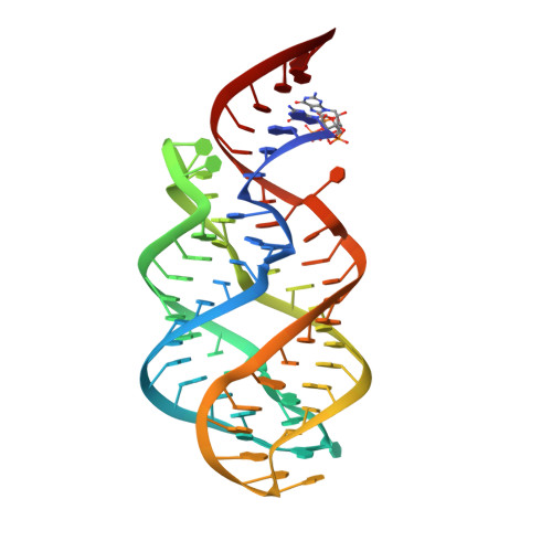
Structural basis for 2'-deoxyguanosine recognition by the 2'-dG-II class of riboswitches.

Matyjasik, M.M.Batey, R.T.

(2019) Nucleic Acids Res 47: 10931-10941

  • DOI: https://doi.org/10.1093/nar/gkz839
  • Primary Citation of Related Structures:  
    6P2H

  • PubMed Abstract: 

    A recent bioinformatic analysis of well-characterized classes of riboswitches uncovered subgroups unable to bind to the regulatory molecule of the parental class. Within the guanine/adenine class, seven groups of RNAs were identified that deviate from the consensus sequence at one or more of three positions directly involved purine nucleobase recognition, one of which was validated as a second class of 2'-deoxyguanosine riboswitch (called 2'-dG-II). To understand how 2'-dG-II riboswitches recognize their cognate ligand and how they differ from a previously identified class of 2'-deoxyguanosine binding riboswitches, we have solved the crystal structure of a 2'-dG-II aptamer domain bound to 2'-deoxyguanosine. This structure reveals a global architecture similar to other members of the purine riboswitch family, but contains key differences within the ligand binding core. Defining the 2'-dG-II riboswitches is a two-nucleotide insertion in the three-way junction that promotes novel base-base interactions. Unlike 2'-dG-I riboswitches, the 2'-dG-II class only requires local changes to the ligand binding pocket of the guanine/adenine class to achieve a change in ligand preference. Notably, members of the 2'-dG-II family have variable ability to discriminate between 2'-deoxyguanosine and riboguanosine, suggesting that a subset of 2'-dG-II riboswitches may bind either molecule to regulate gene expression.


  • Organizational Affiliation

    Department of Biochemistry, University of Colorado at Boulder, Campus Box 596, Boulder, CO 80309-0596, USA.


Macromolecules
Find similar nucleic acids by:  (by identity cutoff)  |  3D Structure
Entity ID: 1
MoleculeChains LengthOrganismImage
RNA (69-MER)69synthetic construct
Sequence Annotations
Expand
  • Reference Sequence
Small Molecules
Ligands 3 Unique
IDChains Name / Formula / InChI Key2D Diagram3D Interactions
GNG (Subject of Investigation/LOI)
Query on GNG

Download Ideal Coordinates CCD File 
B [auth A]2'-DEOXY-GUANOSINE
C10 H13 N5 O4
YKBGVTZYEHREMT-KVQBGUIXSA-N
NCO
Query on NCO

Download Ideal Coordinates CCD File 
C [auth A],
D [auth A],
E [auth A],
F [auth A],
G [auth A]
COBALT HEXAMMINE(III)
Co H18 N6
DYLMFCCYOUSRTK-UHFFFAOYSA-N
MG
Query on MG

Download Ideal Coordinates CCD File 
H [auth A],
I [auth A],
J [auth A],
K [auth A],
L [auth A]
MAGNESIUM ION
Mg
JLVVSXFLKOJNIY-UHFFFAOYSA-N
Experimental Data & Validation

Experimental Data

  • Method: X-RAY DIFFRACTION
  • Resolution: 2.80 Å
  • R-Value Free: 0.276 
  • R-Value Work: 0.244 
  • R-Value Observed: 0.247 
  • Space Group: P 41 2 2
Unit Cell:
Length ( Å )Angle ( ˚ )
a = 91.805α = 90
b = 91.805β = 90
c = 77.756γ = 90
Software Package:
Software NamePurpose
PHENIXrefinement
HKL-3000data reduction
HKL-3000data scaling
PHASERphasing

Structure Validation

View Full Validation Report


Ligand Structure Quality Assessment 


Entry History & Funding Information

Deposition Data


Funding OrganizationLocationGrant Number
National Institutes of Health/National Institute of General Medical Sciences (NIH/NIGMS)United StatesR01-GM073850

Revision History  (Full details and data files)

  • Version 1.0: 2019-10-23
    Type: Initial release
  • Version 1.1: 2019-11-27
    Changes: Database references
  • Version 1.2: 2020-01-01
    Changes: Author supporting evidence
  • Version 1.3: 2023-10-11
    Changes: Data collection, Database references, Derived calculations, Refinement description