6P1L

Crystal structure of EGFR in complex with EAI045


Experimental Data Snapshot

  • Method: X-RAY DIFFRACTION
  • Resolution: 2.80 Å
  • R-Value Free: 0.237 
  • R-Value Work: 0.201 
  • R-Value Observed: 0.203 

wwPDB Validation   3D Report Full Report

Currently 6P1L does not have a validation slider image.


This is version 1.1 of the entry. See complete history


Literature

Discovery and Optimization of Dibenzodiazepinones as Allosteric Mutant-Selective EGFR Inhibitors.

De Clercq, D.J.H.Heppner, D.E.To, C.Jang, J.Park, E.Yun, C.H.Mushajiang, M.Shin, B.H.Gero, T.W.Scott, D.A.Janne, P.A.Eck, M.J.Gray, N.S.

(2019) ACS Med Chem Lett 10: 1549-1553

  • DOI: https://doi.org/10.1021/acsmedchemlett.9b00381
  • Primary Citation of Related Structures:  
    6P1D, 6P1L, 6P8Q

  • PubMed Abstract: 

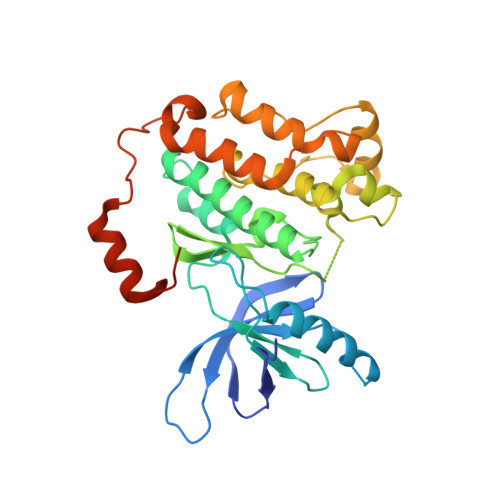
    Allosteric kinase inhibitors represent a promising new therapeutic strategy for targeting kinases harboring oncogenic driver mutations in cancers. Here, we report the discovery, optimization, and structural characterization of allosteric mutant-selective EGFR inhibitors comprising a 5,10-dihydro-11 H -dibenzo[ b , e ][1,4]diazepin-11-one scaffold. Our structure-based medicinal chemistry effort yielded an inhibitor ( 3 ) of the EGFR(L858R/T790M) and EGFR(L858R/T790M/C797S) mutants with an IC 50 of ∼10 nM and high selectivity, as assessed by kinome profiling. Further efforts to develop allosteric dibenzodiazepinone inhibitors may serve as the basis for new therapeutic options for targeting drug-resistant EGFR mutations.


  • Organizational Affiliation

    Department of Cancer Biology, Dana-Farber Cancer Institute, Boston, Massachusetts 02215, United States.


Macromolecules
Find similar proteins by:  (by identity cutoff)  |  3D Structure
Entity ID: 1
MoleculeChains Sequence LengthOrganismDetailsImage
Epidermal growth factor receptor
A, B, C, D
327Homo sapiensMutation(s): 2 
Gene Names: EGFRERBBERBB1HER1
EC: 2.7.10.1
UniProt & NIH Common Fund Data Resources
Find proteins for P00533 (Homo sapiens)
Explore P00533 
Go to UniProtKB:  P00533
PHAROS:  P00533
GTEx:  ENSG00000146648 
Entity Groups  
Sequence Clusters30% Identity50% Identity70% Identity90% Identity95% Identity100% Identity
UniProt GroupP00533
Sequence Annotations
Expand
  • Reference Sequence
Small Molecules
Ligands 3 Unique
IDChains Name / Formula / InChI Key2D Diagram3D Interactions
ANP
Query on ANP

Download Ideal Coordinates CCD File 
F [auth A],
I [auth B],
K [auth C],
N [auth D]
PHOSPHOAMINOPHOSPHONIC ACID-ADENYLATE ESTER
C10 H17 N6 O12 P3
PVKSNHVPLWYQGJ-KQYNXXCUSA-N
9LL (Subject of Investigation/LOI)
Query on 9LL

Download Ideal Coordinates CCD File 
G [auth A],
L [auth C],
O [auth D]
(2R)-2-(5-fluoro-2-hydroxyphenyl)-2-(1-oxo-1,3-dihydro-2H-isoindol-2-yl)-N-(1,3-thiazol-2-yl)acetamide
C19 H14 F N3 O3 S
YTUFHOKUFOQRDF-MRXNPFEDSA-N
MG
Query on MG

Download Ideal Coordinates CCD File 
E [auth A],
H [auth B],
J [auth C],
M [auth D]
MAGNESIUM ION
Mg
JLVVSXFLKOJNIY-UHFFFAOYSA-N
Binding Affinity Annotations 
IDSourceBinding Affinity
9LL BindingDB:  6P1L IC50: min: 3, max: 1000 (nM) from 3 assay(s)
Experimental Data & Validation

Experimental Data

  • Method: X-RAY DIFFRACTION
  • Resolution: 2.80 Å
  • R-Value Free: 0.237 
  • R-Value Work: 0.201 
  • R-Value Observed: 0.203 
  • Space Group: P 1 21 1
Unit Cell:
Length ( Å )Angle ( ˚ )
a = 72.6α = 90
b = 103.15β = 101.18
c = 87.1γ = 90
Software Package:
Software NamePurpose
PHENIXrefinement
Aimlessdata scaling
XDSdata reduction
PHASERphasing

Structure Validation

View Full Validation Report

Currently 6P1L does not have a validation slider image.


Entry History & Funding Information

Deposition Data


Funding OrganizationLocationGrant Number
National Institutes of Health/National Cancer Institute (NIH/NCI)United States--

Revision History  (Full details and data files)

  • Version 1.0: 2019-12-18
    Type: Initial release
  • Version 1.1: 2023-10-11
    Changes: Data collection, Database references, Derived calculations, Refinement description