6P1B

Transcription antitermination factor Q21


Experimental Data Snapshot

  • Method: X-RAY DIFFRACTION
  • Resolution: 1.94 Å
  • R-Value Free: 0.226 
  • R-Value Work: 0.184 
  • R-Value Observed: 0.188 

wwPDB Validation   3D Report Full Report


This is version 1.4 of the entry. See complete history


Literature

Structural basis of Q-dependent antitermination.

Yin, Z.Kaelber, J.T.Ebright, R.H.

(2019) Proc Natl Acad Sci U S A 116: 18384-18390

  • DOI: https://doi.org/10.1073/pnas.1909801116
  • Primary Citation of Related Structures:  
    6P18, 6P19, 6P1A, 6P1B, 6P1C

  • PubMed Abstract: 

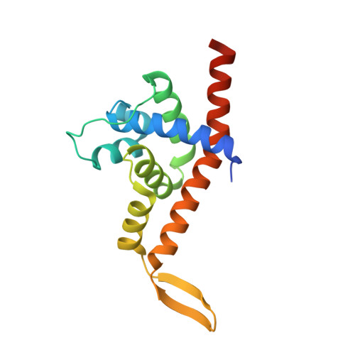
    Lambdoid bacteriophage Q protein mediates the switch from middle to late bacteriophage gene expression by enabling RNA polymerase (RNAP) to read through transcription terminators preceding bacteriophage late genes. Q loads onto RNAP engaged in promoter-proximal pausing at a Q binding element (QBE) and adjacent sigma-dependent pause element (SDPE) to yield a Q-loading complex, and Q subsequently translocates with RNAP as a pausing-deficient, termination-deficient Q-loaded complex. Here, we report high-resolution structures of 4 states on the pathway of antitermination by Q from bacteriophage 21 (Q21): Q21, the Q21-QBE complex, the Q21-loading complex, and the Q21-loaded complex. The results show that Q21 forms a torus, a "nozzle," that narrows and extends the RNAP RNA-exit channel, extruding topologically linked single-stranded RNA and preventing the formation of pause and terminator hairpins.


  • Organizational Affiliation

    Waksman Institute, Rutgers University, Piscataway, NJ 08854.


Macromolecules
Find similar proteins by:  (by identity cutoff)  |  3D Structure
Entity ID: 1
MoleculeChains Sequence LengthOrganismDetailsImage
Q protein
A, B
162Escherichia phage 21Mutation(s): 0 
UniProt
Find proteins for Q9XJQ6 (Escherichia phage 21)
Explore Q9XJQ6 
Go to UniProtKB:  Q9XJQ6
Entity Groups  
Sequence Clusters30% Identity50% Identity70% Identity90% Identity95% Identity100% Identity
UniProt GroupQ9XJQ6
Sequence Annotations
Expand
  • Reference Sequence
Small Molecules
Ligands 2 Unique
IDChains Name / Formula / InChI Key2D Diagram3D Interactions
EDO
Query on EDO

Download Ideal Coordinates CCD File 
G [auth A],
M [auth B],
N [auth B],
O [auth B]
1,2-ETHANEDIOL
C2 H6 O2
LYCAIKOWRPUZTN-UHFFFAOYSA-N
CL
Query on CL

Download Ideal Coordinates CCD File 
C [auth A]
D [auth A]
E [auth A]
F [auth A]
H [auth B]
C [auth A],
D [auth A],
E [auth A],
F [auth A],
H [auth B],
I [auth B],
J [auth B],
K [auth B],
L [auth B]
CHLORIDE ION
Cl
VEXZGXHMUGYJMC-UHFFFAOYSA-M
Experimental Data & Validation

Experimental Data

  • Method: X-RAY DIFFRACTION
  • Resolution: 1.94 Å
  • R-Value Free: 0.226 
  • R-Value Work: 0.184 
  • R-Value Observed: 0.188 
  • Space Group: P 1 21 1
Unit Cell:
Length ( Å )Angle ( ˚ )
a = 32.233α = 90
b = 78.905β = 98.68
c = 68.497γ = 90
Software Package:
Software NamePurpose
DENZOdata reduction
SCALEPACKdata scaling
PHASERphasing
PHENIXrefinement
PDB_EXTRACTdata extraction

Structure Validation

View Full Validation Report


Entry History & Funding Information

Deposition Data


Funding OrganizationLocationGrant Number
National Institutes of Health/National Institute of Environmental Health Sciences (NIH/NIEHS)United StatesGM041376

Revision History  (Full details and data files)

  • Version 1.0: 2019-06-26
    Type: Initial release
  • Version 1.1: 2019-09-11
    Changes: Data collection, Database references
  • Version 1.2: 2019-09-25
    Changes: Data collection, Database references
  • Version 1.3: 2019-12-18
    Changes: Author supporting evidence
  • Version 1.4: 2023-10-11
    Changes: Data collection, Database references, Refinement description