6OY4 | pdb_00006oy4

Crystal structure of complex between recombinant Der p 2.0103 and Fab fragment of 7A1


Experimental Data Snapshot

  • Method: X-RAY DIFFRACTION
  • Resolution: 2.45 Å
  • R-Value Free: 
    0.260 (Depositor), 0.262 (DCC) 
  • R-Value Work: 
    0.203 (Depositor), 0.206 (DCC) 
  • R-Value Observed: 
    0.206 (Depositor) 

Starting Models: experimental
View more details

wwPDB Validation 3D Report Full Report

Validation slider image for 6OY4

This is version 1.5 of the entry. See complete history

Literature

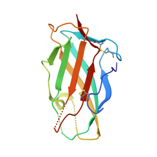
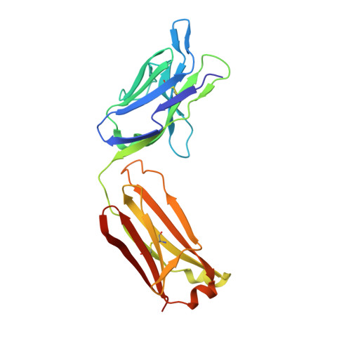
A Human IgE Antibody Binding Site on Der p 2 for the Design of a Recombinant Allergen for Immunotherapy.

Glesner, J.Kapingidza, A.B.Godzwon, M.Offermann, L.R.Mueller, G.A.DeRose, E.F.Wright, P.Richardson, C.M.Woodfolk, J.A.Vailes, L.D.Wunschmann, S.London, R.E.Chapman, M.D.Ohlin, M.Chruszcz, M.Pomes, A.

(2019) J Immunol 203: 2545-2556

  • DOI: https://doi.org/10.4049/jimmunol.1900580
  • Primary Citation Related Structures: 
    6OY4

  • PubMed Abstract: 

    Der p 2 is one of the most important allergens from the house dust mite Dermatophagoides pteronyssinus Identification of human IgE Ab binding epitopes can be used for rational design of allergens with reduced IgE reactivity for therapy. Antigenic analysis of Der p 2 was performed by site-directed mutagenesis based on the x-ray crystal structure of the allergen in complex with a Fab from the murine IgG mAb 7A1 that binds an epitope overlapping with human IgE binding sites. Conformational changes upon Ab binding were confirmed by nuclear magnetic resonance using a 7A1-single-chain variable fragment. In addition, a human IgE Ab construct that interferes with mAb 7A1 binding was isolated from a combinatorial phage-display library constructed from a mite-allergic patient and expressed as two recombinant forms (single-chain Fab in Pichia pastoris and Fab in Escherichia coli ). These two IgE Ab constructs and the mAb 7A1 failed to recognize two Der p 2 epitope double mutants designed to abolish the allergen-Ab interaction while preserving the fold necessary to bind Abs at other sites of the allergen surface. A 10-100-fold reduction in binding of IgE from allergic subjects to the mutants additionally showed that the residues mutated were involved in IgE Ab binding. In summary, mutagenesis of a Der p 2 epitope defined by x-ray crystallography revealed an IgE Ab binding site that will be considered for the design of hypoallergens for immunotherapy.


  • Organizational Affiliation
    • Indoor Biotechnologies, Inc., Charlottesville, VA 22903.

Macromolecule Content 

  • Total Structure Weight: 61.72 kDa 
  • Atom Count: 4,417 
  • Modeled Residue Count: 555 
  • Deposited Residue Count: 566 
  • Unique protein chains: 3

Macromolecules

Find similar proteins by:|  3D Structure
Entity ID: 1
MoleculeChains  Sequence LengthOrganismDetailsImage
Der p 2 variant 3129Dermatophagoides pteronyssinusMutation(s): 0 
UniProt
Find proteins for P49278 (Dermatophagoides pteronyssinus)
Explore P49278 
Go to UniProtKB:  P49278
Entity Groups
Sequence Clusters30% Identity50% Identity70% Identity90% Identity95% Identity100% Identity
UniProt GroupP49278
Sequence Annotations
Expand
Reference Sequence
Find similar proteins by:|  3D Structure
Entity ID: 2
MoleculeChains  Sequence LengthOrganismDetailsImage
Fab fragment of IgG, LIGHT CHAINB [auth C]216Mus musculusMutation(s): 0 
Find similar proteins by:|  3D Structure
Entity ID: 3
MoleculeChains  Sequence LengthOrganismDetailsImage
Fab fragment of IgG, HEAVY CHAINC [auth D]221Mus musculusMutation(s): 0 

Small Molecules

Ligands 1 Unique
IDChains Name / Formula / InChI Key2D Diagram3D Interactions
SO4

Query on SO4


Download:Ideal Coordinates CCD File
D [auth C]SULFATE ION
O4 S
QAOWNCQODCNURD-UHFFFAOYSA-L

Experimental Data & Validation

Experimental Data

  • Method: X-RAY DIFFRACTION
  • Resolution: 2.45 Å
  • R-Value Free:  0.260 (Depositor), 0.262 (DCC) 
  • R-Value Work:  0.203 (Depositor), 0.206 (DCC) 
  • R-Value Observed: 0.206 (Depositor) 
Space Group: C 1 2 1
Unit Cell:
Length ( Å )Angle ( ˚ )
a = 180.094α = 90
b = 43.336β = 125.29
c = 105.935γ = 90
Software Package:
Software NamePurpose
REFMACrefinement
HKL-3000data reduction
HKL-3000data scaling
MOLREPphasing

Structure Validation

View Full Validation Report


Entry History 

& Funding Information

Deposition Data


Funding OrganizationLocationGrant Number
National Institutes of Health/National Institute Of Allergy and Infectious Diseases (NIH/NIAID)United StatesR01AI077653

Revision History  (Full details and data files)

  • Version 1.0: 2019-08-28
    Type: Initial release
  • Version 1.1: 2019-12-18
    Changes: Author supporting evidence
  • Version 1.2: 2020-03-11
    Changes: Database references
  • Version 1.3: 2021-08-04
    Changes: Structure summary
  • Version 1.4: 2023-10-11
    Changes: Data collection, Database references, Refinement description
  • Version 1.5: 2024-10-16
    Changes: Structure summary