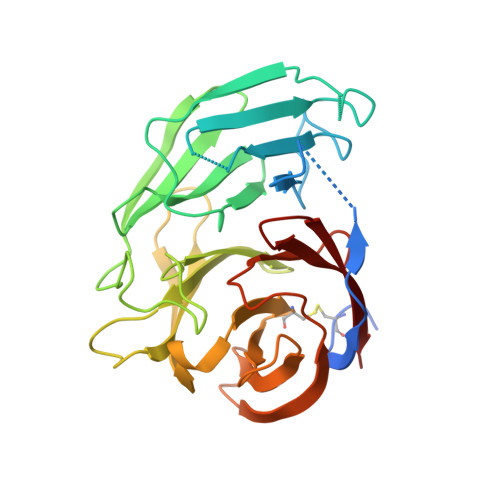
Stable calcium-free myocilin olfactomedin domain variants reveal challenges in differentiating between benign and glaucoma-causing mutations.
Hill, S.E., Kwon, M.S., Martin, M.D., Suntharalingam, A., Hazel, A., Dickey, C.A., Gumbart, J.C., Lieberman, R.L.(2019) J Biol Chem 294: 12717-12728
- PubMed: 31270212 Search on PubMedSearch on PubMed Central
- DOI: https://doi.org/10.1074/jbc.RA119.009419
- Primary Citation Related Structures:
6OU0, 6OU1, 6OU2, 6OU3 - PubMed Abstract:
Nonsynonymous gene mutations can be beneficial, neutral, or detrimental to the stability, structure, and biological function of the encoded protein, but the effects of these mutations are often not readily predictable. For example, the β-propeller olfactomedin domain of myocilin (mOLF) exhibits a complex interrelationship among structure(s), stability, and aggregation. Numerous mutations within mOLF are linked to glaucoma; the resulting variants are less stable, aggregation-prone, and sequestered intracellularly, causing cytotoxicity. Here, we report the first stable mOLF variants carrying substitutions in the calcium-binding site that exhibit solution characteristics indistinguishable from those of glaucoma variants. Crystal structures of these stable variants at 1.8-2.0-Å resolution revealed features that we could not predict by molecular dynamics simulations, including loss of loop structure, helix unwinding, and a blade shift. Double mutants that combined a stabilizing substitution and a selected glaucoma-causing single-point mutant rescued in vitro folding and stability defects. In the context of full-length myocilin, secretion of stable single variants was indistinguishable from that of the WT protein, and the double mutants were secreted to varying extents. In summary, our finding that mOLF can tolerate particular substitutions that render the protein stable despite a conformational switch emphasizes the complexities in differentiating between benign and glaucoma-causing variants and provides new insight into the possible biological function of myocilin.
- School of Chemistry and Biochemistry, Georgia Institute of Technology, Atlanta, Georgia 30332.
Organizational Affiliation: