Functional divergence caused by mutations in an energetic hotspot in ERK2.
Taylor 4th, C.A., Cormier, K.W., Keenan, S.E., Earnest, S., Stippec, S., Wichaidit, C., Juang, Y.C., Wang, J., Shvartsman, S.Y., Goldsmith, E.J., Cobb, M.H.(2019) Proc Natl Acad Sci U S A 116: 15514-15523
- PubMed: 31296562 Search on PubMedSearch on PubMed Central
- DOI: https://doi.org/10.1073/pnas.1905015116
- Primary Citation Related Structures:
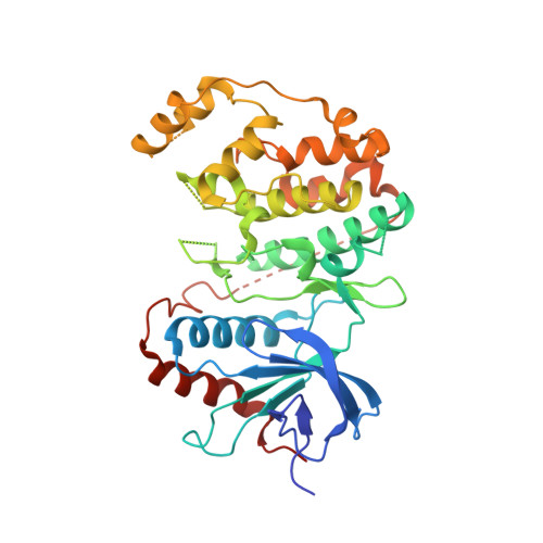
6OT6, 6OTS - PubMed Abstract:
The most frequent extracellular signal-regulated kinase 2 (ERK2) mutation occurring in cancers is E322K (E-K). ERK2 E-K reverses a buried charge in the ERK2 common docking (CD) site, a region that binds activators, inhibitors, and substrates. Little is known about the cellular consequences associated with this mutation, other than apparent increases in tumor resistance to pathway inhibitors. ERK2 E-K, like the mutation of the preceding aspartate (ERK2 D321N [D-N]) known as the sevenmaker mutation, causes increased activity in cells and evades inactivation by dual-specificity phosphatases. As opposed to findings in cancer cells, in developmental assays in Drosophila , only ERK2 D-N displays a significant gain of function, revealing mutation-specific phenotypes. The crystal structure of ERK2 D-N is indistinguishable from that of wild-type protein, yet this mutant displays increased thermal stability. In contrast, the crystal structure of ERK2 E-K reveals profound structural changes, including disorder in the CD site and exposure of the activation loop phosphorylation sites, which likely account for the decreased thermal stability of the protein. These contiguous mutations in the CD site of ERK2 are both required for docking interactions but lead to unpredictably different functional outcomes. Our results suggest that the CD site is in an energetically strained configuration, and this helps drive conformational changes at distal sites on ERK2 during docking interactions.
- Department of Pharmacology, UT Southwestern Medical Center, Dallas, TX 75390.
Organizational Affiliation: