6OQQ

Legionella pneumophila SidJ/Saccharomyces cerevisiae calmodulin complex


Experimental Data Snapshot

  • Method: X-RAY DIFFRACTION
  • Resolution: 2.10 Å
  • R-Value Free: 0.234 
  • R-Value Work: 0.202 
  • R-Value Observed: 0.203 

wwPDB Validation   3D Report Full Report


Ligand Structure Quality Assessment 


This is version 1.3 of the entry. See complete history


Literature

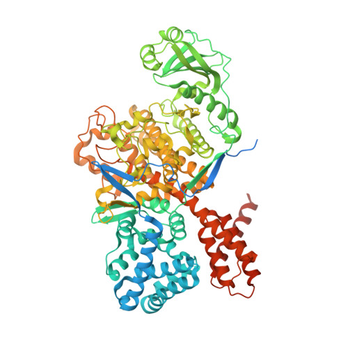
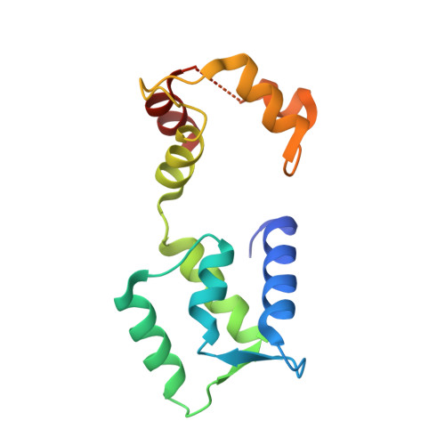
Bacterial pseudokinase catalyzes protein polyglutamylation to inhibit the SidE-family ubiquitin ligases.

Black, M.H.Osinski, A.Gradowski, M.Servage, K.A.Pawlowski, K.Tomchick, D.R.Tagliabracci, V.S.

(2019) Science 364: 787-792

  • DOI: https://doi.org/10.1126/science.aaw7446
  • Primary Citation of Related Structures:  
    6OQQ

  • PubMed Abstract: 

    Enzymes with a protein kinase fold transfer phosphate from adenosine 5'-triphosphate (ATP) to substrates in a process known as phosphorylation. Here, we show that the Legionella meta-effector SidJ adopts a protein kinase fold, yet unexpectedly catalyzes protein polyglutamylation. SidJ is activated by host-cell calmodulin to polyglutamylate the SidE family of ubiquitin (Ub) ligases. Crystal structures of the SidJ-calmodulin complex reveal a protein kinase fold that catalyzes ATP-dependent isopeptide bond formation between the amino group of free glutamate and the γ-carboxyl group of an active-site glutamate in SidE. We show that SidJ polyglutamylation of SidE, and the consequent inactivation of Ub ligase activity, is required for successful Legionella replication in a viable eukaryotic host cell.


  • Organizational Affiliation

    Department of Molecular Biology, University of Texas Southwestern Medical Center, Dallas, TX 75390, USA.


Macromolecules
Find similar proteins by:  (by identity cutoff)  |  3D Structure
Entity ID: 1
MoleculeChains Sequence LengthOrganismDetailsImage
Legionella pneumophila SidJ
A, C
873Legionella pneumophilaMutation(s): 0 
Gene Names: SidJ
UniProt
Find proteins for Q5ZTK6 (Legionella pneumophila subsp. pneumophila (strain Philadelphia 1 / ATCC 33152 / DSM 7513))
Explore Q5ZTK6 
Go to UniProtKB:  Q5ZTK6
Entity Groups  
Sequence Clusters30% Identity50% Identity70% Identity90% Identity95% Identity100% Identity
UniProt GroupQ5ZTK6
Sequence Annotations
Expand
  • Reference Sequence
Find similar proteins by:  (by identity cutoff)  |  3D Structure
Entity ID: 2
MoleculeChains Sequence LengthOrganismDetailsImage
Calmodulin
B, D
147Saccharomyces cerevisiae S288CMutation(s): 0 
Gene Names: CMD1YBR109CYBR0904
UniProt
Find proteins for P06787 (Saccharomyces cerevisiae (strain ATCC 204508 / S288c))
Explore P06787 
Go to UniProtKB:  P06787
Entity Groups  
Sequence Clusters30% Identity50% Identity70% Identity90% Identity95% Identity100% Identity
UniProt GroupP06787
Sequence Annotations
Expand
  • Reference Sequence
Small Molecules
Ligands 6 Unique
IDChains Name / Formula / InChI Key2D Diagram3D Interactions
AMP
Query on AMP

Download Ideal Coordinates CCD File 
E [auth A],
X [auth C]
ADENOSINE MONOPHOSPHATE
C10 H14 N5 O7 P
UDMBCSSLTHHNCD-KQYNXXCUSA-N
POP
Query on POP

Download Ideal Coordinates CCD File 
F [auth A],
Y [auth C],
Z [auth C]
PYROPHOSPHATE 2-
H2 O7 P2
XPPKVPWEQAFLFU-UHFFFAOYSA-L
EDO
Query on EDO

Download Ideal Coordinates CCD File 
DA [auth C]
EA [auth C]
FA [auth C]
GA [auth C]
L [auth A]
DA [auth C],
EA [auth C],
FA [auth C],
GA [auth C],
L [auth A],
M [auth A],
N [auth A],
O [auth A],
P [auth A],
Q [auth A],
R [auth A],
S [auth A],
T [auth A]
1,2-ETHANEDIOL
C2 H6 O2
LYCAIKOWRPUZTN-UHFFFAOYSA-N
CA
Query on CA

Download Ideal Coordinates CCD File 
HA [auth D],
IA [auth D],
U [auth B],
V [auth B]
CALCIUM ION
Ca
BHPQYMZQTOCNFJ-UHFFFAOYSA-N
MG
Query on MG

Download Ideal Coordinates CCD File 
AA [auth C]
BA [auth C]
CA [auth C]
G [auth A]
H [auth A]
AA [auth C],
BA [auth C],
CA [auth C],
G [auth A],
H [auth A],
I [auth A],
J [auth A]
MAGNESIUM ION
Mg
JLVVSXFLKOJNIY-UHFFFAOYSA-N
NA
Query on NA

Download Ideal Coordinates CCD File 
K [auth A],
W [auth B]
SODIUM ION
Na
FKNQFGJONOIPTF-UHFFFAOYSA-N
Experimental Data & Validation

Experimental Data

  • Method: X-RAY DIFFRACTION
  • Resolution: 2.10 Å
  • R-Value Free: 0.234 
  • R-Value Work: 0.202 
  • R-Value Observed: 0.203 
  • Space Group: P 1 21 1
Unit Cell:
Length ( Å )Angle ( ˚ )
a = 105.49α = 90
b = 104.749β = 103.97
c = 107.77γ = 90
Software Package:
Software NamePurpose
PHENIXrefinement
HKL-3000data scaling
PDB_EXTRACTdata extraction
HKL-3000data reduction
MLPHAREphasing

Structure Validation

View Full Validation Report


Ligand Structure Quality Assessment 


Entry History & Funding Information

Deposition Data


Funding OrganizationLocationGrant Number
National Institutes of Health/National Institute of General Medical Sciences (NIH/NIGMS)United StatesT32GM008014-34
National Institutes of Health/National Heart, Lung, and Blood Institute (NIH/NHLBI)United StatesF30HL143859-01
National Institutes of Health/National Institute of Diabetes and Digestive and Kidney Disease (NIH/NIDDK)United StatesR00DK099254
National Institutes of Health/National Institute of General Medical Sciences (NIH/NIGMS)United StatesT32GM-7062-44
Robert A. Welch FoundationUnited StatesI-1911
Other governmentUnited StatesRP170674
Polish National Science CentrePoland2014/15/B/NZ1/03559

Revision History  (Full details and data files)

  • Version 1.0: 2019-05-15
    Type: Initial release
  • Version 1.1: 2019-06-12
    Changes: Data collection, Database references
  • Version 1.2: 2019-12-04
    Changes: Author supporting evidence
  • Version 1.3: 2024-03-13
    Changes: Data collection, Database references, Derived calculations