6OPM | pdb_00006opm

Casposase bound to integration product


Experimental Data Snapshot

  • Method: X-RAY DIFFRACTION
  • Resolution: 3.10 Å
  • R-Value Free: 
    0.256 (Depositor), 0.280 (DCC) 
  • R-Value Work: 
    0.198 (Depositor), 0.213 (DCC) 
  • R-Value Observed: 
    0.200 (Depositor) 

wwPDB Validation 3D Report Full Report

Validation slider image for 6OPM

This is version 1.2 of the entry. See complete history

Literature

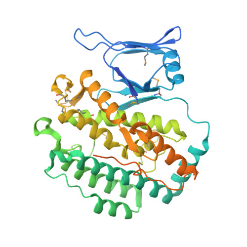
Casposase structure and the mechanistic link between DNA transposition and spacer acquisition by CRISPR-Cas.

Hickman, A.B.Kailasan, S.Genzor, P.Haase, A.D.Dyda, F.

(2020) Elife 9

  • DOI: https://doi.org/10.7554/eLife.50004
  • Primary Citation Related Structures: 
    6OPM

  • PubMed Abstract: 

    Key to CRISPR-Cas adaptive immunity is maintaining an ongoing record of invading nucleic acids, a process carried out by the Cas1-Cas2 complex that integrates short segments of foreign genetic material (spacers) into the CRISPR locus. It is hypothesized that Cas1 evolved from casposases, a novel class of transposases. We show here that the Methanosarcina mazei casposase can integrate varied forms of the casposon end in vitro, and recapitulates several properties of CRISPR-Cas integrases including site-specificity. The X-ray structure of the casposase bound to DNA representing the product of integration reveals a tetramer with target DNA bound snugly between two dimers in which single-stranded casposon end binding resembles that of spacer 3'-overhangs. The differences between transposase and CRISPR-Cas integrase are largely architectural, and it appears that evolutionary change involved changes in protein-protein interactions to favor Cas2 binding over tetramerization; this in turn led to preferred integration of single spacers over two transposon ends.


  • Organizational Affiliation
    • Laboratory of Molecular Biology, National Institute of Diabetes and Digestive and Kidney Diseases, National Institutes of Health, Bethesda, United States.

Macromolecule Content 

  • Total Structure Weight: 215.1 kDa 
  • Atom Count: 12,315 
  • Modeled Residue Count: 1,463 
  • Deposited Residue Count: 1,792 
  • Unique protein chains: 2
  • Unique nucleic acid chains: 1

Macromolecules


Find similar proteins by:|  3D Structure
Entity ID: 1
MoleculeChains  Sequence LengthOrganismDetailsImage
CRISPR-associated endonuclease Cas1
A, B, C, D
431Methanosarcina mazeiMutation(s): 1 
Gene Names: 
EC: 3.1
UniProt
Find proteins for A0A0F8IEL4 (Methanosarcina mazei)
Explore A0A0F8IEL4 
Go to UniProtKB:  A0A0F8IEL4
Entity Groups
Sequence Clusters30% Identity50% Identity70% Identity90% Identity95% Identity100% Identity
UniProt GroupA0A0F8IEL4
Sequence Annotations
Expand
Reference Sequence
Find similar proteins by:  Sequence   |   3D Structure  
Entity ID: 3
MoleculeChains  Sequence LengthOrganismDetailsImage
unknownG [auth H],
H [auth J]
13Methanosarcina mazeiMutation(s): 0 
Find similar nucleic acids by:  Sequence
Entity ID: 2
MoleculeChains LengthOrganismImage
DNA 21-mer
E, F
21Methanosarcina mazei
Sequence Annotations
Expand
Reference Sequence

Experimental Data & Validation

Experimental Data

  • Method: X-RAY DIFFRACTION
  • Resolution: 3.10 Å
  • R-Value Free:  0.256 (Depositor), 0.280 (DCC) 
  • R-Value Work:  0.198 (Depositor), 0.213 (DCC) 
  • R-Value Observed: 0.200 (Depositor) 
Space Group: P 43 21 2
Unit Cell:
Length ( Å )Angle ( ˚ )
a = 106.39α = 90
b = 106.39β = 90
c = 423.36γ = 90
Software Package:
Software NamePurpose
BUSTERrefinement
XDSdata reduction
XSCALEdata scaling
SHARPphasing

Structure Validation

View Full Validation Report


Entry History 

Deposition Data

Revision History  (Full details and data files)

  • Version 1.0: 2020-02-12
    Type: Initial release
  • Version 1.1: 2020-08-26
    Changes: Database references, Derived calculations
  • Version 1.2: 2024-11-20
    Changes: Data collection, Database references, Structure summary