Targetable HER3 functions driving tumorigenic signaling in HER2-amplified cancers.
Campbell, M.R., Ruiz-Saenz, A., Peterson, E., Agnew, C., Ayaz, P., Garfinkle, S., Littlefield, P., Steri, V., Oeffinger, J., Sampang, M., Shan, Y., Shaw, D.E., Jura, N., Moasser, M.M.(2022) Cell Rep 38: 110291-110291
- PubMed: 35108525 Search on PubMedSearch on PubMed Central
- DOI: https://doi.org/10.1016/j.celrep.2021.110291
- Primary Citation Related Structures:
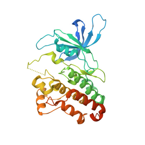
6OP9 - PubMed Abstract:
Effective inactivation of the HER2-HER3 tumor driver has remained elusive because of the challenging attributes of the pseudokinase HER3. We report a structure-function study of constitutive HER2-HER3 signaling to identify opportunities for targeting. The allosteric activation of the HER2 kinase domain (KD) by the HER3 KD is required for tumorigenic signaling and can potentially be targeted by allosteric inhibitors. ATP binding within the catalytically inactive HER3 KD provides structural rigidity that is important for signaling, but this is mimicked, not opposed, by small molecule ATP analogs, reported here in a bosutinib-bound crystal structure. Mutational disruption of ATP binding and molecular dynamics simulation of the apo KD of HER3 identify a conformational coupling of the ATP pocket with a hydrophobic AP-2 pocket, analogous to EGFR, that is critical for tumorigenic signaling and feasible for targeting. The value of these potential target sites is confirmed in tumor growth assays using gene replacement techniques.
- Department of Medicine, University of California, San Francisco, San Francisco, CA 94143, USA.
Organizational Affiliation: