A Single Salt Bridge in VIM-20 Increases Protein Stability and Antibiotic Resistance under Low-Zinc Conditions.
Cheng, Z., Shurina, B.A., Bethel, C.R., Thomas, P.W., Marshall, S.H., Thomas, C.A., Yang, K., Kimble, R.L., Montgomery, J.S., Orischak, M.G., Miller, C.M., Tennenbaum, J.L., Nix, J.C., Tierney, D.L., Fast, W., Bonomo, R.A., Page, R.C., Crowder, M.W.(2019) mBio 10
- PubMed: 31744917 Search on PubMedSearch on PubMed Central
- DOI: https://doi.org/10.1128/mBio.02412-19
- Primary Citation Related Structures:
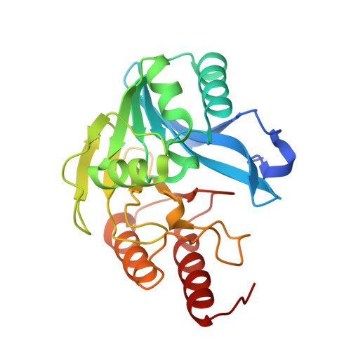
6OP6, 6OP7 - PubMed Abstract:
To understand the evolution of Verona integron-encoded metallo-β-lactamase (VIM) genes ( bla VIM ) and their clinical impact, microbiological, biochemical, and structural studies were conducted. Forty-five clinically derived VIM variants engineered in a uniform background and expressed in Escherichia coli afforded increased resistance toward all tested antibiotics; the variants belonging to the VIM-1-like and VIM-4-like families exhibited higher MICs toward five out of six antibiotics than did variants belonging to the widely distributed and clinically important VIM-2-like family. Generally, maximal MIC increases were observed when cephalothin and imipenem were tested. Additionally, MIC determinations under conditions with low zinc availability suggested that some VIM variants are also evolving to overcome zinc deprivation. The most profound increase in resistance was observed in VIM-2-like variants (e.g., VIM-20 H229R) at low zinc availability. Biochemical analyses reveal that VIM-2 and VIM-20 exhibited similar metal binding properties and steady-state kinetic parameters under the conditions tested. Crystal structures of VIM-20 in the reduced and oxidized forms at 1.25 Å and 1.37 Å resolution, respectively, show that Arg229 forms an additional salt bridge with Glu171. Differential scanning fluorimetry of purified proteins and immunoblots of periplasmic extracts revealed that this difference increases thermostability and resistance to proteolytic degradation when zinc availability is low. Therefore, zinc scarcity appears to be a selective pressure driving the evolution of multiple metallo-β-lactamase families, although compensating mutations use different mechanisms to enhance resistance. IMPORTANCE Antibiotic resistance is a growing clinical threat. One of the most serious areas of concern is the ability of some bacteria to degrade carbapenems, drugs that are often reserved as last-resort antibiotics. Resistance to carbapenems can be conferred by a large group of related enzymes called metallo-β-lactamases that rely on zinc ions for function and for overall stability. Here, we studied an extensive panel of 45 different metallo-β-lactamases from a subfamily called VIM to discover what changes are emerging as resistance evolves in clinical settings. Enhanced resistance to some antibiotics was observed. We also found that at least one VIM variant developed a new ability to remain more stable under conditions where zinc availability is limited, and we determined the origin of this stability in atomic detail. These results suggest that zinc scarcity helps drive the evolution of this resistance determinant.
- Department of Chemistry and Biochemistry, Miami University, Oxford, Ohio, USA.
Organizational Affiliation: