6OOU

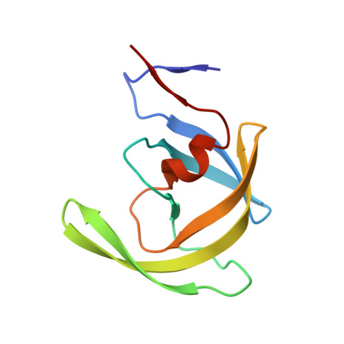
Crystal structure of HIV-1 Protease NL4-3 L89V Mutant in complex with darunavir


Experimental Data Snapshot

  • Method: X-RAY DIFFRACTION
  • Resolution: 2.13 Å
  • R-Value Free: 0.255 
  • R-Value Work: 0.204 
  • R-Value Observed: 0.206 

Starting Model: experimental
View more details

wwPDB Validation   3D Report Full Report

Currently 6OOU does not have a validation slider image.


This is version 1.3 of the entry. See complete history


Literature

Molecular Determinants of Epistasis in HIV-1 Protease: Elucidating the Interdependence of L89V and L90M Mutations in Resistance.

Henes, M.Kosovrasti, K.Lockbaum, G.J.Leidner, F.Nachum, G.S.Nalivaika, E.A.Bolon, D.N.A.Kurt Yilmaz, N.Schiffer, C.A.Whitfield, T.W.

(2019) Biochemistry 58: 3711-3726

  • DOI: https://doi.org/10.1021/acs.biochem.9b00446
  • Primary Citation of Related Structures:  
    6OOS, 6OOT, 6OOU

  • PubMed Abstract: 

    Protease inhibitors have the highest potency among antiviral therapies against HIV-1 infections, yet the virus can evolve resistance. Darunavir (DRV), currently the most potent Food and Drug Administration-approved protease inhibitor, retains potency against single-site mutations. However, complex combinations of mutations can confer resistance to DRV. While the interdependence between mutations within HIV-1 protease is key for inhibitor potency, the molecular mechanisms that underlie this control remain largely unknown. In this study, we investigated the interdependence between the L89V and L90M mutations and their effects on DRV binding. These two mutations have been reported to be positively correlated with one another in HIV-1 patient-derived protease isolates, with the presence of one mutation making the probability of the occurrence of the second mutation more likely. The focus of our investigation is a patient-derived isolate, with 24 mutations that we call "KY"; this variant includes the L89V and L90M mutations. Three additional KY variants with back-mutations, KY(V89L), KY(M90L), and the KY(V89L/M90L) double mutation, were used to experimentally assess the individual and combined effects of these mutations on DRV inhibition and substrate processing. The enzymatic assays revealed that the KY(V89L) variant, with methionine at residue 90, is highly resistant, but its catalytic function is compromised. When a leucine to valine mutation at residue 89 is present simultaneously with the L90M mutation, a rescue of catalytic efficiency is observed. Molecular dynamics simulations of these DRV-bound protease variants reveal how the L90M mutation induces structural changes throughout the enzyme that undermine the binding interactions.


Macromolecules
Find similar proteins by:  (by identity cutoff)  |  3D Structure
Entity ID: 1
MoleculeChains Sequence LengthOrganismDetailsImage
NL4-3 PROTEASEA [auth B],
B [auth A]
99Human immunodeficiency virus 1Mutation(s): 2 
Gene Names: pol
UniProt
Find proteins for P12497 (Human immunodeficiency virus type 1 group M subtype B (isolate NY5))
Explore P12497 
Go to UniProtKB:  P12497
Entity Groups  
Sequence Clusters30% Identity50% Identity70% Identity90% Identity95% Identity100% Identity
UniProt GroupP12497
Sequence Annotations
Expand
  • Reference Sequence
Small Molecules
Ligands 2 Unique
IDChains Name / Formula / InChI Key2D Diagram3D Interactions
017
Query on 017

Download Ideal Coordinates CCD File 
C [auth A](3R,3AS,6AR)-HEXAHYDROFURO[2,3-B]FURAN-3-YL(1S,2R)-3-[[(4-AMINOPHENYL)SULFONYL](ISOBUTYL)AMINO]-1-BENZYL-2-HYDROXYPROPYLCARBAMATE
C27 H37 N3 O7 S
CJBJHOAVZSMMDJ-HEXNFIEUSA-N
SO4
Query on SO4

Download Ideal Coordinates CCD File 
D [auth A],
E [auth A]
SULFATE ION
O4 S
QAOWNCQODCNURD-UHFFFAOYSA-L
Binding Affinity Annotations 
IDSourceBinding Affinity
017 BindingDB:  6OOU Ki: min: 5.00e-3, max: 0.25 (nM) from 2 assay(s)
Experimental Data & Validation

Experimental Data

  • Method: X-RAY DIFFRACTION
  • Resolution: 2.13 Å
  • R-Value Free: 0.255 
  • R-Value Work: 0.204 
  • R-Value Observed: 0.206 
  • Space Group: P 21 21 21
Unit Cell:
Length ( Å )Angle ( ˚ )
a = 51.298α = 90
b = 58.27β = 90
c = 62.444γ = 90
Software Package:
Software NamePurpose
PHENIXrefinement
HKL-3000data scaling
PHASERphasing
Cootmodel building

Structure Validation

View Full Validation Report

Currently 6OOU does not have a validation slider image.


Entry History & Funding Information

Deposition Data


Funding OrganizationLocationGrant Number
National Institutes of Health/National Institute of General Medical Sciences (NIH/NIGMS)United StatesP01-GM109767

Revision History  (Full details and data files)

  • Version 1.0: 2019-08-21
    Type: Initial release
  • Version 1.1: 2019-09-11
    Changes: Data collection, Database references
  • Version 1.2: 2020-01-01
    Changes: Author supporting evidence
  • Version 1.3: 2023-10-11
    Changes: Data collection, Database references, Refinement description, Structure summary