6OOI | pdb_00006ooi

Crystal structure of triosephosphate isomerase from Schistosoma mansoni in complex with 2PG


Experimental Data Snapshot

  • Method: X-RAY DIFFRACTION
  • Resolution: 2.14 Å
  • R-Value Free: 
    0.229 (Depositor), 0.229 (DCC) 
  • R-Value Work: 
    0.180 (Depositor), 0.180 (DCC) 
  • R-Value Observed: 
    0.181 (Depositor) 

Starting Model: experimental
View more details

wwPDB Validation 3D Report Full Report

Validation slider image for 6OOI

Ligand Structure Quality Assessment 


This is version 1.2 of the entry. See complete history

Literature

Crystal structures of Triosephosphate Isomerases from Taenia solium and Schistosoma mansoni provide insights for vaccine rationale and drug design against helminth parasites.

Jimenez-Sandoval, P.Castro-Torres, E.Gonzalez-Gonzalez, R.Diaz-Quezada, C.Gurrola, M.Camacho-Manriquez, L.D.Leyva-Navarro, L.Brieba, L.G.

(2020) PLoS Negl Trop Dis 14: e0007815-e0007815

  • DOI: https://doi.org/10.1371/journal.pntd.0007815
  • Primary Citation Related Structures: 
    6OOG, 6OOI

  • PubMed Abstract: 

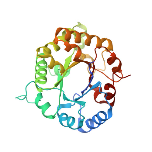
    Triosephosphate isomerases (TPIs) from Taenia solium (TsTPI) and Schistosoma mansoni (SmTPI) are potential vaccine and drug targets against cysticercosis and schistosomiasis, respectively. This is due to the dependence of parasitic helminths on glycolysis and because those proteins elicit an immune response, presumably due to their surface localization. Here we report the crystal structures of TsTPI and SmTPI in complex with 2-phosphoglyceric acid (2-PGA). Both TPIs fold into a dimeric (β-α)8 barrel in which the dimer interface consists of α-helices 2, 3, and 4, and swapping of loop 3. TPIs from parasitic helminths harbor a region of three amino acids knows as the SXD/E insert (S155 to E157 and S157 to D159 in TsTPI and SmTPI, respectively). This insert is located between α5 and β6 and is proposed to be the main TPI epitope. This region is part of a solvent-exposed 310-helix that folds into a hook-like structure. The crystal structures of TsTPI and SmTPI predicted conformational epitopes that could be used for vaccine design. Surprisingly, the epitopes corresponding to the SXD/E inserts are not the ones with the greatest immunological potential. SmTPI, but not TsTPI, habors a sole solvent exposed cysteine (SmTPI-S230) and alterations in this residue decrease catalysis. The latter suggests that thiol-conjugating agents could be used to target SmTPI. In sum, the crystal structures of SmTPI and TsTPI are a blueprint for targeted schistosomiasis and cysticercosis drug and vaccine development.


  • Organizational Affiliation
    • Laboratorio Nacional de Genómica para la Biodiversidad, Centro de Investigación y de Estudios Avanzados del IPN, Irapuato, Guanajuato, México.

Macromolecule Content 

  • Total Structure Weight: 229.48 kDa 
  • Atom Count: 16,312 
  • Modeled Residue Count: 2,003 
  • Deposited Residue Count: 2,048 
  • Unique protein chains: 1

Macromolecules

Find similar proteins by:|  3D Structure
Entity ID: 1
MoleculeChains  Sequence LengthOrganismDetailsImage
Triosephosphate isomerase
A, B, C, D, E
A, B, C, D, E, F, G, H
256Schistosoma mansoniMutation(s): 0 
Gene Names: TPI
EC: 5.3.1.1
UniProt
Find proteins for P48501 (Schistosoma mansoni)
Explore P48501 
Go to UniProtKB:  P48501
Entity Groups
Sequence Clusters30% Identity50% Identity70% Identity90% Identity95% Identity100% Identity
UniProt GroupP48501
Sequence Annotations
Expand
Reference Sequence

Small Molecules

Ligands 4 Unique
IDChains Name / Formula / InChI Key2D Diagram3D Interactions
PGA

Query on PGA


Download:Ideal Coordinates CCD File
BA [auth G]
EA [auth H]
I [auth A]
M [auth B]
Q [auth C]
BA [auth G],
EA [auth H],
I [auth A],
M [auth B],
Q [auth C],
U [auth D],
W [auth E],
Z [auth F]
2-PHOSPHOGLYCOLIC ACID
C2 H5 O6 P
ASCFNMCAHFUBCO-UHFFFAOYSA-N
GOL

Query on GOL


Download:Ideal Coordinates CCD File
DA [auth H],
P [auth C]
GLYCEROL
C3 H8 O3
PEDCQBHIVMGVHV-UHFFFAOYSA-N
CL

Query on CL


Download:Ideal Coordinates CCD File
AA [auth F]
CA [auth G]
J [auth A]
N [auth B]
R [auth C]
AA [auth F],
CA [auth G],
J [auth A],
N [auth B],
R [auth C],
V [auth D]
CHLORIDE ION
Cl
VEXZGXHMUGYJMC-UHFFFAOYSA-M
NA

Query on NA


Download:Ideal Coordinates CCD File
FA [auth H]
K [auth A]
L [auth A]
O [auth B]
S [auth C]
FA [auth H],
K [auth A],
L [auth A],
O [auth B],
S [auth C],
T [auth C],
X [auth E],
Y [auth E]
SODIUM ION
Na
FKNQFGJONOIPTF-UHFFFAOYSA-N

Experimental Data & Validation

Experimental Data

  • Method: X-RAY DIFFRACTION
  • Resolution: 2.14 Å
  • R-Value Free:  0.229 (Depositor), 0.229 (DCC) 
  • R-Value Work:  0.180 (Depositor), 0.180 (DCC) 
  • R-Value Observed: 0.181 (Depositor) 
Space Group: P 43
Unit Cell:
Length ( Å )Angle ( ˚ )
a = 108.05α = 90
b = 108.05β = 90
c = 182.72γ = 90
Software Package:
Software NamePurpose
PHENIXrefinement
CrystalCleardata collection
Cootmodel building
Aimlessdata reduction

Structure Validation

View Full Validation Report


Ligand Structure Quality Assessment 


Entry History 

& Funding Information

Deposition Data


Funding OrganizationLocationGrant Number
Consejo Nacional de Ciencia y Tecnologia (CONACYT)MexicoFronteras de la Ciencia 13

Revision History  (Full details and data files)

  • Version 1.0: 2020-01-08
    Type: Initial release
  • Version 1.1: 2020-01-22
    Changes: Database references
  • Version 1.2: 2023-10-11
    Changes: Data collection, Database references, Refinement description