The multifunctional globin dehaloperoxidase strikes again: Simultaneous peroxidase and peroxygenase mechanisms in the oxidation of EPA pollutants.
Malewschik, T., de Serrano, V., McGuire, A.H., Ghiladi, R.A.(2019) Arch Biochem Biophys 673: 108079-108079
- PubMed: 31445024 Search on PubMed
- DOI: https://doi.org/10.1016/j.abb.2019.108079
- Primary Citation Related Structures:
6ONG, 6ONK, 6ONR, 6ONX, 6ONZ, 6OO1, 6OO6, 6OO8 - PubMed Abstract:
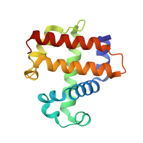
The multifunctional catalytic hemoglobin dehaloperoxidase (DHP) from the terebellid polychaete Amphitrite ornata was found to catalyze the H 2 O 2 -dependent oxidation of EPA Priority Pollutants (4-Me-o-cresol, 4-Cl-m-cresol and pentachlorophenol) and EPA Toxic Substances Control Act compounds (o-, m-, p-cresol and 4-Cl-o-cresol). Biochemical assays (HPLC/LC-MS) indicated formation of multiple oxidation products, including the corresponding catechol, 2-methylbenzoquinone (2-MeBq), and oligomers with varying degrees of oxidation and/or dehalogenation. Using 4-Br-o-cresol as a representative substrate, labeling studies with 18 O confirmed that the O-atom incorporated into the catechol was derived exclusively from H 2 O 2 , whereas the O-atom incorporated into 2-MeBq was from H 2 O, consistent with this single substrate being oxidized by both peroxygenase and peroxidase mechanisms, respectively. Stopped-flow UV-visible spectroscopic studies strongly implicate a role for Compound I in the peroxygenase mechanism leading to catechol formation, and for Compounds I and ES in the peroxidase mechanism that yields the 2-MeBq product. The X-ray crystal structures of DHP bound with 4-F-o-cresol (1.42 Å; PDB 6ONG), 4-Cl-o-cresol (1.50 Å; PDB 6ONK), 4-Br-o-cresol (1.70 Å; PDB 6ONX), 4-NO 2 -o-cresol (1.80 Å; PDB 6ONZ), o-cresol (1.60 Å; PDB 6OO1), p-cresol (2.10 Å; PDB 6OO6), 4-Me-o-cresol (1.35 Å; PDB 6ONR) and pentachlorophenol (1.80 Å; PDB 6OO8) revealed substrate binding sites in the distal pocket in close proximity to the heme cofactor, consistent with both oxidation mechanisms. The findings establish cresols as a new class of substrate for DHP, demonstrate that multiple oxidation mechanisms may exist for a given substrate, and provide further evidence that different substituents can serve as functional switches between the different activities performed by dehaloperoxidase. More broadly, the results demonstrate the complexities of marine pollution where both microbial and non-microbial systems may play significant roles in the biotransformations of EPA-classified pollutants, and further reinforces that heterocyclic compounds of anthropogenic origin should be considered as environmental stressors of infaunal organisms.
- Department of Chemistry, North Carolina State University, Raleigh, NC, 27695-8204, USA.
Organizational Affiliation: