Biochemical and Structural Characterization of XoxG and XoxJ and Their Roles in Lanthanide-Dependent Methanol Dehydrogenase Activity.
Featherston, E.R., Rose, H.R., McBride, M.J., Taylor, E.M., Boal, A.K., Cotruvo Jr., J.A.(2019) Chembiochem 20: 2360-2372
- PubMed: 31017712 Search on PubMedSearch on PubMed Central
- DOI: https://doi.org/10.1002/cbic.201900184
- Primary Citation Related Structures:
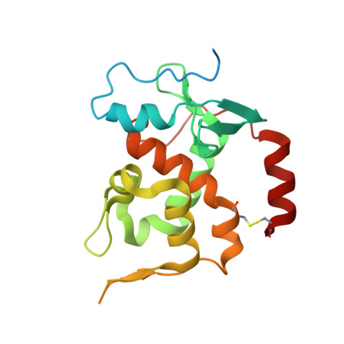
6ONP, 6ONQ - PubMed Abstract:
Lanthanide (Ln)-dependent methanol dehydrogenases (MDHs) have recently been shown to be widespread in methylotrophic bacteria. Along with the core MDH protein, XoxF, these systems contain two other proteins, XoxG (a c-type cytochrome) and XoxJ (a periplasmic binding protein of unknown function), about which little is known. In this work, we have biochemically and structurally characterized these proteins from the methyltroph Methylobacterium extorquens AM1. In contrast to results obtained in an artificial assay system, assays of XoxFs metallated with La III , Ce III , and Nd III using their physiological electron acceptor, XoxG, display Ln-independent activities, but the K m for XoxG markedly increases from La to Nd. This result suggests that XoxG's redox properties are tuned specifically for lighter Lns in XoxF, an interpretation supported by the unusually low reduction potential of XoxG (+172 mV). The X-ray crystal structure of XoxG provides a structural basis for this reduction potential and insight into the XoxG-XoxF interaction. Finally, the X-ray crystal structure of XoxJ reveals a large hydrophobic cleft and suggests a role in the activation of XoxF. These studies enrich our understanding of the underlying chemical principles that enable the activity of XoxF with multiple lanthanides in vitro and in vivo.
- Department of Chemistry, The Pennsylvania State University, University Park, PA, 16802, USA.
Organizational Affiliation: