


Crystal structure of 8-amino-7-oxononanoate synthase from Burkholderia phymatum
Conrady, D.G., Abendroth, J., Lorimer, D.D., Horanyi, P.S., Edwards, T.E.To be published.
Experimental Data Snapshot
Starting Model: experimental
View more details
wwPDB Validation 3D Report Full Report

Entity ID: 1 | |||||
|---|---|---|---|---|---|
| Molecule | Chains | Sequence Length | Organism | Details | Image |
| 8-amino-7-oxononanoate synthase | 402 | Paraburkholderia phymatum STM815 | Mutation(s): 0 Gene Names: bioF, Bphy_0172 EC: 2.3.1.47 | 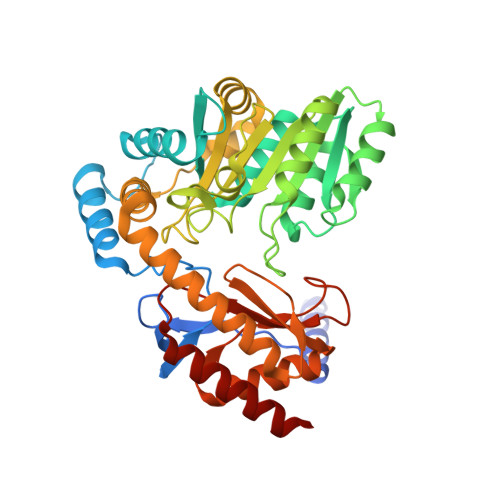 | |
UniProt | |||||
Entity Groups | |||||
| Sequence Clusters | 30% Identity50% Identity70% Identity90% Identity95% Identity100% Identity | ||||
| UniProt Group | B2JKH6 | ||||
Sequence AnnotationsExpand | |||||
Reference Sequence | |||||
| Length ( Å ) | Angle ( ˚ ) |
|---|---|
| a = 78.7 | α = 90 |
| b = 92.58 | β = 90 |
| c = 125.56 | γ = 90 |
| Software Name | Purpose |
|---|---|
| PHENIX | refinement |
| XDS | data reduction |
| XSCALE | data scaling |
| PDB_EXTRACT | data extraction |
| MOLREP | phasing |


