6OMQ | pdb_00006omq

Crystal structure of PtmU3 complexed with PTM substrate


Experimental Data Snapshot

  • Method: X-RAY DIFFRACTION
  • Resolution: 1.86 Å
  • R-Value Free: 
    0.235 (Depositor), 0.243 (DCC) 
  • R-Value Work: 
    0.181 (Depositor), 0.191 (DCC) 
  • R-Value Observed: 
    0.184 (Depositor) 

Starting Model: experimental
View more details

wwPDB Validation 3D Report Full Report

Validation slider image for 6OMQ

Ligand Structure Quality Assessment 


This is version 1.4 of the entry. See complete history

Literature

Characterization and Crystal Structure of a Nonheme Diiron Monooxygenase Involved in Platensimycin and Platencin Biosynthesis.

Dong, L.B.Liu, Y.C.Cepeda, A.J.Kalkreuter, E.Deng, M.R.Rudolf, J.D.Chang, C.Joachimiak, A.Phillips Jr., G.N.Shen, B.

(2019) J Am Chem Soc 141: 12406-12412

  • DOI: https://doi.org/10.1021/jacs.9b06183
  • Primary Citation Related Structures: 
    6OMP, 6OMQ, 6OMR

  • PubMed Abstract: 

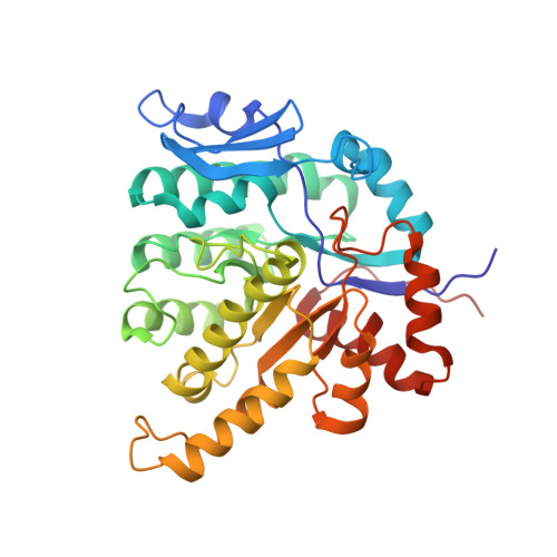
    Nonheme diiron monooxygenases make up a rapidly growing family of oxygenases that are rarely identified in secondary metabolism. Herein, we report the in vivo, in vitro, and structural characterizations of a nonheme diiron monooxygenase, PtmU3, that installs a C-5 β-hydroxyl group in the unified biosynthesis of platensimycin and platencin, two highly functionalized diterpenoids that act as potent and selective inhibitors of bacterial and mammalian fatty acid synthases. This hydroxylation sets the stage for the subsequent A-ring cleavage step key to the unique diterpene-derived scaffolds of platensimycin and platencin. PtmU3 adopts an unprecedented triosephosphate isomerase (TIM) barrel structural fold for this class of enzymes and possesses a noncanonical diiron active site architecture with a saturated six-coordinate iron center lacking a μ-oxo bridge. This study reveals the first member of a previously unidentified superfamily of TIM-barrel-fold enzymes for metal-dependent dioxygen activation, with the majority predicted to act on CoA-linked substrates, thus expanding our knowledge of nature's repertoire of nonheme diiron monooxygenases and TIM-barrel-fold enzymes.


  • Organizational Affiliation
    • Midwest Center for Structural Genomics and Structural Biology Center, Biosciences Division , Argonne National Laboratory , Argonne , Illinois 60439 , United States.

Macromolecule Content 

  • Total Structure Weight: 80.42 kDa 
  • Atom Count: 6,482 
  • Modeled Residue Count: 712 
  • Deposited Residue Count: 712 
  • Unique protein chains: 1

Macromolecules

Find similar proteins by:|  3D Structure
Entity ID: 1
MoleculeChains  Sequence LengthOrganismDetailsImage
PtmU3
A, B
356Streptomyces platensisMutation(s): 0 
UniProt
Find proteins for A0A0A0UVH9 (Streptomyces platensis)
Explore A0A0A0UVH9 
Go to UniProtKB:  A0A0A0UVH9
Entity Groups
Sequence Clusters30% Identity50% Identity70% Identity90% Identity95% Identity100% Identity
UniProt GroupA0A0A0UVH9
Sequence Annotations
Expand
Reference Sequence

Small Molecules

Ligands 3 Unique
IDChains Name / Formula / InChI Key2D Diagram3D Interactions
MZD

Query on MZD


Download:Ideal Coordinates CCD File
O [auth B]S-[2-({N-[(2R)-2-hydroxy-3,3-dimethyl-4-(phosphonooxy)butanoyl]-beta-alanyl}amino)ethyl] (5beta,7alpha,8alpha,10alpha,11alpha,13alpha)-7-hydroxy-11,16-epoxykaurane-18-thioate
C31 H51 N2 O10 P S
WNJOABVUVJTUCG-HDNPFTGKSA-N
ACT

Query on ACT


Download:Ideal Coordinates CCD File
C [auth A]
D [auth A]
E [auth A]
F [auth A]
G [auth A]
C [auth A],
D [auth A],
E [auth A],
F [auth A],
G [auth A],
K [auth B],
L [auth B],
M [auth B],
N [auth B]
ACETATE ION
C2 H3 O2
QTBSBXVTEAMEQO-UHFFFAOYSA-M
MN

Query on MN


Download:Ideal Coordinates CCD File
H [auth A]
I [auth A]
J [auth A]
P [auth B]
Q [auth B]
H [auth A],
I [auth A],
J [auth A],
P [auth B],
Q [auth B],
R [auth B],
S [auth B]
MANGANESE (II) ION
Mn
WAEMQWOKJMHJLA-UHFFFAOYSA-N

Experimental Data & Validation

Experimental Data

  • Method: X-RAY DIFFRACTION
  • Resolution: 1.86 Å
  • R-Value Free:  0.235 (Depositor), 0.243 (DCC) 
  • R-Value Work:  0.181 (Depositor), 0.191 (DCC) 
  • R-Value Observed: 0.184 (Depositor) 
Space Group: I 2 2 2
Unit Cell:
Length ( Å )Angle ( ˚ )
a = 112.564α = 90
b = 122.864β = 90
c = 139.936γ = 90
Software Package:
Software NamePurpose
HKL-2000data reduction
SCALEPACKdata scaling
PHASERphasing
REFMACrefinement
PDB_EXTRACTdata extraction

Structure Validation

View Full Validation Report


Ligand Structure Quality Assessment 


Entry History 

& Funding Information

Deposition Data


Funding OrganizationLocationGrant Number
National Institutes of Health/National Institute of General Medical Sciences (NIH/NIGMS)United StatesGM114353

Revision History  (Full details and data files)

  • Version 1.0: 2019-07-24
    Type: Initial release
  • Version 1.1: 2019-07-31
    Changes: Data collection, Database references
  • Version 1.2: 2019-08-21
    Changes: Data collection, Database references
  • Version 1.3: 2020-01-01
    Changes: Author supporting evidence
  • Version 1.4: 2023-10-11
    Changes: Data collection, Database references, Derived calculations, Refinement description