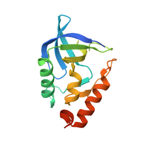
Toward the computational design of protein crystals with improved resolution.
Jeliazkov, J.R., Robinson, A.C., Garcia-Moreno E, B., Berger, J.M., Gray, J.J.(2019) Acta Crystallogr D Struct Biol 75: 1015-1027
- PubMed: 31692475
- DOI: https://doi.org/10.1107/S2059798319013226
- Primary Citation of Related Structures:
6OK8, 6U0W, 6U0X - PubMed Abstract:
Substantial advances have been made in the computational design of protein interfaces over the last 20 years. However, the interfaces targeted by design have typically been stable and high-affinity. Here, we report the development of a generic computational design method to stabilize the weak interactions at crystallographic interfaces. Initially, we analyzed structures reported in the Protein Data Bank to determine whether crystals with more stable interfaces result in higher resolution structures. We found that for 22 variants of a single protein crystallized by a single individual, the Rosetta-calculated `crystal score' correlates with the reported diffraction resolution. We next developed and tested a computational design protocol, seeking to identify point mutations that would improve resolution in a highly stable variant of staphylococcal nuclease (SNase). Using a protocol based on fixed protein backbones, only one of the 11 initial designs crystallized, indicating modeling inaccuracies and forcing us to re-evaluate our strategy. To compensate for slight changes in the local backbone and side-chain environment, we subsequently designed on an ensemble of minimally perturbed protein backbones. Using this strategy, four of the seven designed proteins crystallized. By collecting diffraction data from multiple crystals per design and solving crystal structures, we found that the designed crystals improved the resolution modestly and in unpredictable ways, including altering the crystal space group. Post hoc, in silico analysis of the three observed space groups for SNase showed that the native space group was the lowest scoring for four of six variants (including the wild type), but that resolution did not correlate with crystal score, as it did in the preliminary results. Collectively, our results show that calculated crystal scores can correlate with reported resolution, but that the correlation is absent when the problem is inverted. This outcome suggests that more comprehensive modeling of the crystallographic state is necessary to design high-resolution protein crystals from poorly diffracting crystals.
- Program in Molecular Biophysics, Johns Hopkins University, Baltimore, Maryland, USA.
Organizational Affiliation:


