6OIL

Crystal structure of human VISTA extracellular domain


Experimental Data Snapshot

  • Method: X-RAY DIFFRACTION
  • Resolution: 1.85 Å
  • R-Value Free: 0.223 
  • R-Value Work: 0.180 
  • R-Value Observed: 0.182 

wwPDB Validation   3D Report Full Report


Ligand Structure Quality Assessment 


This is version 1.5 of the entry. See complete history


Literature

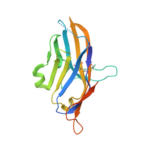
Structure and Functional Binding Epitope of V-domain Ig Suppressor of T Cell Activation.

Mehta, N.Maddineni, S.Mathews, I.I.Andres Parra Sperberg, R.Huang, P.S.Cochran, J.R.

(2019) Cell Rep 28: 2509-2516.e5

  • DOI: https://doi.org/10.1016/j.celrep.2019.07.073
  • Primary Citation of Related Structures:  
    6OIL

  • PubMed Abstract: 

    V-domain immunoglobulin (Ig) suppressor of T cell activation (VISTA) is an immune checkpoint protein that inhibits the T cell response against cancer. Similar to PD-1 and CTLA-4, a blockade of VISTA promotes tumor clearance by the immune system. Here, we report a 1.85 Å crystal structure of the elusive human VISTA extracellular domain, whose lack of homology necessitated a combinatorial MR-Rosetta approach for structure determination. We highlight features that make the VISTA immunoglobulin variable (IgV)-like fold unique among B7 family members, including two additional disulfide bonds and an extended loop region with an attached helix that we show forms a contiguous binding epitope for a clinically relevant anti-VISTA antibody. We propose an overlap of this antibody-binding region with the binding epitope for V-set and Ig domain containing 3 (VSIG3), a purported functional binding partner of VISTA. The structure and functional epitope presented here will help guide future drug development efforts against this important checkpoint target.


  • Organizational Affiliation

    Department of Bioengineering, Stanford University, Stanford, CA 94305, USA.


Macromolecules
Find similar proteins by:  (by identity cutoff)  |  3D Structure
Entity ID: 1
MoleculeChains Sequence LengthOrganismDetailsImage
V-type immunoglobulin domain-containing suppressor of T-cell activation168Homo sapiensMutation(s): 4 
Gene Names: VSIR
UniProt & NIH Common Fund Data Resources
Find proteins for Q9H7M9 (Homo sapiens)
Explore Q9H7M9 
Go to UniProtKB:  Q9H7M9
PHAROS:  Q9H7M9
GTEx:  ENSG00000107738 
Entity Groups  
Sequence Clusters30% Identity50% Identity70% Identity90% Identity95% Identity100% Identity
UniProt GroupQ9H7M9
Sequence Annotations
Expand
  • Reference Sequence
Small Molecules
Ligands 1 Unique
IDChains Name / Formula / InChI Key2D Diagram3D Interactions
NAG
Query on NAG

Download Ideal Coordinates CCD File 
B [auth A],
C [auth A]
2-acetamido-2-deoxy-beta-D-glucopyranose
C8 H15 N O6
OVRNDRQMDRJTHS-FMDGEEDCSA-N
Experimental Data & Validation

Experimental Data

  • Method: X-RAY DIFFRACTION
  • Resolution: 1.85 Å
  • R-Value Free: 0.223 
  • R-Value Work: 0.180 
  • R-Value Observed: 0.182 
  • Space Group: P 1 21 1
Unit Cell:
Length ( Å )Angle ( ˚ )
a = 29.169α = 90
b = 64.089β = 91.73
c = 37.695γ = 90
Software Package:
Software NamePurpose
XDSdata reduction
XSCALEdata scaling
PHENIXrefinement
PDB_EXTRACTdata extraction
PHENIXphasing

Structure Validation

View Full Validation Report


Ligand Structure Quality Assessment 


Entry History & Funding Information

Deposition Data


Funding OrganizationLocationGrant Number
Department of Energy (DOE, United States)United StatesDE-AC02-76SF00515
National Institutes of Health/National Institute of General Medical Sciences (NIH/NIGMS)United StatesP41GM103393

Revision History  (Full details and data files)

  • Version 1.0: 2019-09-11
    Type: Initial release
  • Version 1.1: 2019-09-18
    Changes: Data collection, Structure summary
  • Version 1.2: 2019-11-13
    Changes: Database references
  • Version 1.3: 2019-12-04
    Changes: Author supporting evidence
  • Version 1.4: 2020-07-29
    Type: Remediation
    Reason: Carbohydrate remediation
    Changes: Data collection, Derived calculations, Structure summary
  • Version 1.5: 2024-04-03
    Changes: Data collection, Database references, Refinement description, Structure summary